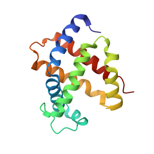
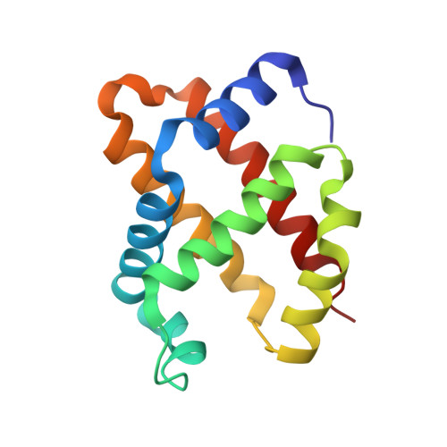
The functional similarity and structural diversity of human and cartilaginous fish hemoglobins.
Naoi, Y., Chong, K.T., Yoshimatsu, K., Miyazaki, G., Tame, J.R., Park, S.Y., Adachi, S., Morimoto, H.(2001) J Mol Biology 307: 259-270
- PubMed: 11243818
- DOI: https://doi.org/10.1006/jmbi.2000.4446
- Primary Citation of Related Structures:
1GCV, 1GCW - PubMed Abstract:
Although many descriptions of adaptive molecular evolution of vertebrate hemoglobins (Hb) can be found in physiological text books, they are based mainly on changes of the primary structure and place more emphasis on conservation than alterations at the functional site. Sequence analysis alone, however, does not reveal much about the evolution of new functions in proteins. It was found recently that there are many functionally important structural differences between human and a ray (Dasyatis akajei) Hb even where sequence is conserved between the two. We have solved the structures of the deoxy and CO forms of a second cartilaginous fish (a shark, Mustelus griseus) Hb, and compared it with structures of human Hb, two bony fish Hbs and the ray Hb in order to understand more about how vertebrate Hbs have functionally evolved by the selection of random amino acid substitutions. The sequence identity of cartilaginous fish Hb and human Hb is a little less than 40 %, with many functionally important amino acid replacements. Wider substitutions than usually considered as neutral have been accepted in the course of molecular evolution of Hb. As with the ray Hb, the shark Hb shows functionally important structural differences from human Hb that involve amino acid substitutions and shifts of preserved amino acid residues induced by substitutions in other parts of the molecule. Most importantly, beta E11Val in deoxy human Hb, which overlaps the ligand binding site and is considered to play a key role in controlling the oxygen affinity, moves away about 1 A in both the shark and ray Hbs. Thus adaptive molecular evolution is feasible as a result of both functionally significant mutations and deviations of preserved amino acid residues induced by other amino acid substitutions.
Organizational Affiliation:
Division of Biophysical Engineering, Graduate School of Engineering Science, Osaka University, Toyonaka, Osaka 560-8531, Japan. naoi@food2.kyoto-u.ac.jp