1GGC

MAJOR ANTIGEN-INDUCED DOMAIN REARRANGEMENTS IN AN ANTIBODY


Experimental Data Snapshot

  • Method: X-RAY DIFFRACTION
  • Resolution: 2.80 Å
  • R-Value Work: 0.190 
  • R-Value Observed: 0.190 

wwPDB Validation   3D Report Full Report


This is version 1.2 of the entry. See complete history


Literature

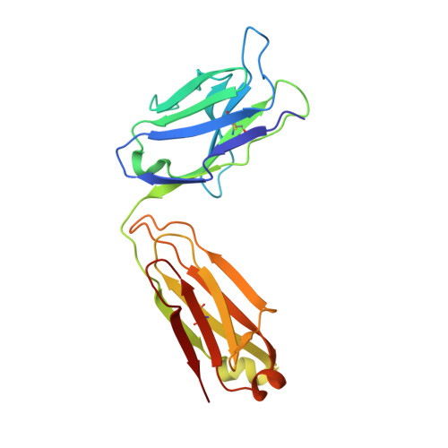
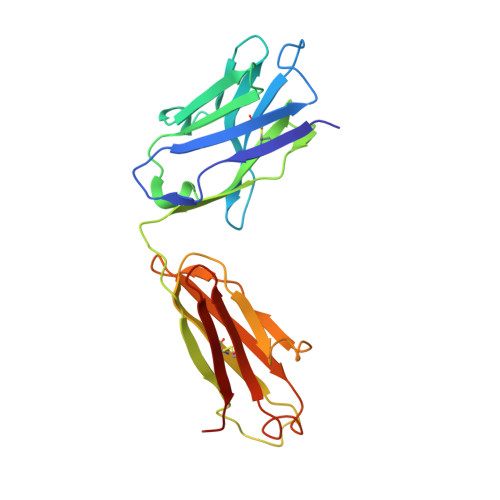
Major antigen-induced domain rearrangements in an antibody.

Stanfield, R.L.Takimoto-Kamimura, M.Rini, J.M.Profy, A.T.Wilson, I.A.

(1993) Structure 1: 83-93

  • DOI: https://doi.org/10.1016/0969-2126(93)90024-b
  • Primary Citation of Related Structures:  
    1GGB, 1GGC

  • PubMed Abstract: 

    Recent structural results have shown that antibodies use an induced fit mechanism to recognize and bind their antigens. Here we present the crystallographically determined structure of an Fab directed against an HIV-1 peptide (Fab 50.1) in the unliganded state and compare it with the peptide-bound structure. We perform a detailed analysis of the components that contribute to enhanced antigen binding and recognition. Induced fit of Fab 50.1 to its peptide antigen involves a substantial rearrangement of the third complementarity determining region loop of the heavy chain (H3), as well as a large rotation of the variable heavy (VH) chain relative to the variable light (VL) chain. Analysis of other Fab structures suggests that the extent of the surface area buried at the VL-VH interface correlates with the ability to alter antibody quaternary structure by reorientation of the VL-VH domains. Fab 50.1 exhibits the largest conformational changes yet observed in a single antibody. These can be attributed to the flexibility of the variable region. Comparisons of new data with previous examples lend to the general conclusion that a small VL-VH interface, due in part to a short H3 loop, permits substantial alterations to the antigen-binding pocket. This has major implications for the prediction, engineering and design of antibody-combining sites.


  • Organizational Affiliation

    Department of Molecular Biology, Scripps Research Institute, La Jolla, CA 92037.


Macromolecules
Find similar proteins by:  (by identity cutoff)  |  3D Structure
Entity ID: 1
MoleculeChains Sequence LengthOrganismDetailsImage
IGG2A-KAPPA 50.1 FAB (LIGHT CHAIN)A [auth L]215Mus musculusMutation(s): 0 
Entity Groups  
Sequence Clusters30% Identity50% Identity70% Identity90% Identity95% Identity100% Identity
Sequence Annotations
Expand
  • Reference Sequence
Find similar proteins by:  (by identity cutoff)  |  3D Structure
Entity ID: 2
MoleculeChains Sequence LengthOrganismDetailsImage
IGG2A-KAPPA 50.1 FAB (HEAVY CHAIN)B [auth H]215Mus musculusMutation(s): 0 
UniProt
Find proteins for P01865 (Mus musculus)
Explore P01865 
Go to UniProtKB:  P01865
Entity Groups  
Sequence Clusters30% Identity50% Identity70% Identity90% Identity95% Identity100% Identity
UniProt GroupP01865
Sequence Annotations
Expand
  • Reference Sequence
Experimental Data & Validation

Experimental Data

  • Method: X-RAY DIFFRACTION
  • Resolution: 2.80 Å
  • R-Value Work: 0.190 
  • R-Value Observed: 0.190 
  • Space Group: P 21 21 21
Unit Cell:
Length ( Å )Angle ( ˚ )
a = 83.7α = 90
b = 110.7β = 90
c = 56.3γ = 90
Software Package:
Software NamePurpose
X-PLORmodel building
X-PLORrefinement
X-PLORphasing

Structure Validation

View Full Validation Report


Entry History 

Deposition Data

Revision History  (Full details and data files)

  • Version 1.0: 1993-10-31
    Type: Initial release
  • Version 1.1: 2008-03-24
    Changes: Version format compliance
  • Version 1.2: 2011-07-13
    Changes: Version format compliance