4LSW

Crystallization and Structural Analysis of 2-Hydroxyacid Dehydrogenase from Ketogulonicigenium vulgare Y25


Experimental Data Snapshot

  • Method: X-RAY DIFFRACTION
  • Resolution: 1.64 Å
  • R-Value Free: 0.251 
  • R-Value Work: 0.211 
  • R-Value Observed: 0.213 

wwPDB Validation   3D Report Full Report


This is version 1.2 of the entry. See complete history


Literature

Crystallization and structural analysis of 2-hydroxyacid dehydrogenase from Ketogulonicigenium vulgare.

Han, X.Xiong, X.Hu, X.Li, M.Zhang, W.Liu, X.

(2014) Biotechnol Lett 36: 295-300

  • DOI: https://doi.org/10.1007/s10529-013-1354-8
  • Primary Citation of Related Structures:  
    4LSW

  • PubMed Abstract: 

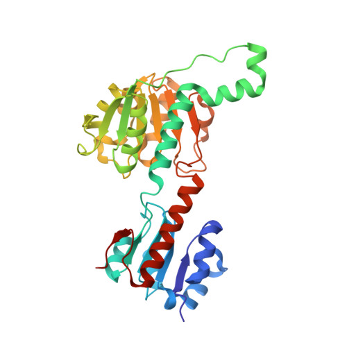
    L-2-Hydroxyacid dehydrogenase (HDH) from Ketogulonicigenium vulgare Y25 was cloned and overexpressed in Escherichia coli. The protein was purified and crystallized by the sitting-drop vapour-diffusion method with polyethylene glycol 3350 as precipitant. The crystal structure of HDH was determined at 1.64 Å resolution using the molecular replacement method with the crystal structure of hydroxyl (phenyl) pyruvate reductase from Coleus blumei Benth as the search model. The overall structure of HDH was similar to that of hydroxyl(phenyl)pyruvate reductase, consisting of two compact domains separated by a deep active cleft. The most significant structural divergence is located around the pocket gate comprising residues A210, T211 and R212, which is located on top of the catalytic triad.


  • Organizational Affiliation

    State Key Laboratory of Medicinal Chemical Biology, College of Life Sciences, Nankai University, Tianjin, 300071, China, hxdon@126.com.


Macromolecules
Find similar proteins by:  (by identity cutoff)  |  3D Structure
Entity ID: 1
MoleculeChains Sequence LengthOrganismDetailsImage
D-2-hydroxyacid dehydrogensase protein318Ketogulonicigenium vulgare Y25Mutation(s): 0 
Entity Groups  
Sequence Clusters30% Identity50% Identity70% Identity90% Identity95% Identity100% Identity
Sequence Annotations
Expand
  • Reference Sequence
Experimental Data & Validation

Experimental Data

  • Method: X-RAY DIFFRACTION
  • Resolution: 1.64 Å
  • R-Value Free: 0.251 
  • R-Value Work: 0.211 
  • R-Value Observed: 0.213 
  • Space Group: C 1 2 1
Unit Cell:
Length ( Å )Angle ( ˚ )
a = 111.202α = 90
b = 52.333β = 107.46
c = 65.647γ = 90
Software Package:
Software NamePurpose
HKL-2000data collection
PHASESphasing
PHENIXrefinement
HKL-2000data reduction
HKL-2000data scaling

Structure Validation

View Full Validation Report


Entry History 

Deposition Data

Revision History  (Full details and data files)

  • Version 1.0: 2013-09-11
    Type: Initial release
  • Version 1.1: 2015-06-17
    Changes: Database references
  • Version 1.2: 2023-11-08
    Changes: Data collection, Database references, Refinement description