6SMN

A. thaliana serine hydroxymethyltransferase isoform 2 (AtSHMT2) in complex with methotrexate


Experimental Data Snapshot

  • Method: X-RAY DIFFRACTION
  • Resolution: 1.63 Å
  • R-Value Free: 0.177 
  • R-Value Work: 0.125 

wwPDB Validation   3D Report Full Report


Ligand Structure Quality Assessment 


This is version 1.1 of the entry. See complete history


Literature

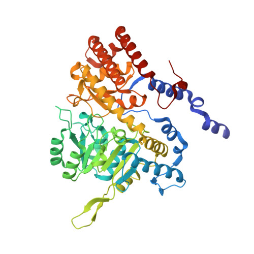
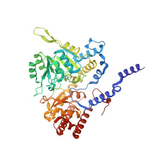
Structural basis of methotrexate and pemetrexed action on serine hydroxymethyltransferases revealed using plant models.

Ruszkowski, M.Sekula, B.Ruszkowska, A.Contestabile, R.Nogues, I.Angelaccio, S.Szczepaniak, A.Dauter, Z.

(2019) Sci Rep 9: 19614-19614

  • DOI: https://doi.org/10.1038/s41598-019-56043-4
  • Primary Citation of Related Structures:  
    6SMN, 6SMR, 6SMW

  • PubMed Abstract: 

    Serine hydroxymethyltransferases (SHMTs) reversibly transform serine into glycine in a reaction accompanied with conversion of tetrahydrofolate (THF) into 5,10-methylene-THF (5,10-meTHF). In vivo, 5,10-meTHF is the main carrier of one-carbon (1C) units, which are utilized for nucleotide biosynthesis and other processes crucial for every living cell, but hyperactivated in overproliferating cells (e.g. cancer tissues). SHMTs are emerging as a promising target for development of new drugs because it appears possible to inhibit growth of cancer cells by cutting off the supply of 5,10-meTHF. Methotrexate (MTX) and pemetrexed (PTX) are two examples of antifolates that have cured many patients over the years but target different enzymes from the folate cycle (mainly dihydrofolate reductase and thymidylate synthase, respectively). Here we show crystal structures of MTX and PTX bound to plant SHMT isozymes from cytosol and mitochondria-human isozymes exist in the same subcellular compartments. We verify inhibition of the studied isozymes by a thorough kinetic analysis. We propose to further exploit antifolate scaffold in development of SHMT inhibitors because it seems likely that especially polyglutamylated PTX inhibits SHMTs in vivo. Structure-based optimization is expected to yield novel antifolates that could potentially be used as chemotherapeutics.


  • Organizational Affiliation

    Synchrotron Radiation Research Section of MCL, National Cancer Institute, Argonne, IL, USA. mruszkowski@ibch.poznan.pl.


Macromolecules
Find similar proteins by:  (by identity cutoff)  |  3D Structure
Entity ID: 1
MoleculeChains Sequence LengthOrganismDetailsImage
Serine hydroxymethyltransferase 2, mitochondrial
A, B, C
480Arabidopsis thalianaMutation(s): 0 
Gene Names: SHM2SHMT2At5g26780F2P16.40
EC: 2.1.2.1
UniProt
Find proteins for Q94C74 (Arabidopsis thaliana)
Explore Q94C74 
Go to UniProtKB:  Q94C74
Entity Groups  
Sequence Clusters30% Identity50% Identity70% Identity90% Identity95% Identity100% Identity
UniProt GroupQ94C74
Sequence Annotations
Expand
  • Reference Sequence
Find similar proteins by:  (by identity cutoff)  |  3D Structure
Entity ID: 2
MoleculeChains Sequence LengthOrganismDetailsImage
Serine hydroxymethyltransferase 2, mitochondrial480Arabidopsis thalianaMutation(s): 0 
Gene Names: SHM2SHMT2At5g26780F2P16.40
EC: 2.1.2.1
UniProt
Find proteins for Q94C74 (Arabidopsis thaliana)
Explore Q94C74 
Go to UniProtKB:  Q94C74
Entity Groups  
Sequence Clusters30% Identity50% Identity70% Identity90% Identity95% Identity100% Identity
UniProt GroupQ94C74
Sequence Annotations
Expand
  • Reference Sequence
Small Molecules
Ligands 4 Unique
IDChains Name / Formula / InChI Key2D Diagram3D Interactions
MTX (Subject of Investigation/LOI)
Query on MTX

Download Ideal Coordinates CCD File 
G [auth A],
JA [auth C],
XA [auth C],
Y [auth B]
METHOTREXATE
C20 H22 N8 O5
FBOZXECLQNJBKD-ZDUSSCGKSA-N
PLS
Query on PLS

Download Ideal Coordinates CCD File 
E [auth A],
HA [auth C],
W [auth B]
[3-HYDROXY-2-METHYL-5-PHOSPHONOOXYMETHYL-PYRIDIN-4-YLMETHYL]-SERINE
C11 H17 N2 O8 P
ODVKKQWXKRZJLG-VIFPVBQESA-N
SER
Query on SER

Download Ideal Coordinates CCD File 
AB [auth D]SERINE
C3 H7 N O3
MTCFGRXMJLQNBG-REOHCLBHSA-N
EDO
Query on EDO

Download Ideal Coordinates CCD File 
AA [auth B]
BA [auth B]
BB [auth D]
CA [auth B]
CB [auth D]
AA [auth B],
BA [auth B],
BB [auth D],
CA [auth B],
CB [auth D],
DA [auth B],
DB [auth D],
EA [auth B],
EB [auth D],
F [auth A],
FA [auth B],
FB [auth D],
GA [auth B],
GB [auth D],
H [auth A],
HB [auth D],
I [auth A],
IA [auth C],
J [auth A],
K [auth A],
KA [auth C],
L [auth A],
LA [auth C],
M [auth A],
MA [auth C],
N [auth A],
NA [auth C],
O [auth A],
OA [auth C],
P [auth A],
PA [auth C],
Q [auth A],
QA [auth C],
R [auth A],
RA [auth C],
S [auth A],
SA [auth C],
T [auth A],
TA [auth C],
U [auth A],
UA [auth C],
V [auth B],
VA [auth C],
WA [auth C],
X [auth B],
YA [auth C],
Z [auth B],
ZA [auth D]
1,2-ETHANEDIOL
C2 H6 O2
LYCAIKOWRPUZTN-UHFFFAOYSA-N
Modified Residues  1 Unique
IDChains TypeFormula2D DiagramParent
LLP
Query on LLP
D
L-PEPTIDE LINKINGC14 H22 N3 O7 PLYS
Binding Affinity Annotations 
IDSourceBinding Affinity
MTX Binding MOAD:  6SMN Ki: 2.70e+5 (nM) from 1 assay(s)
Experimental Data & Validation

Experimental Data

  • Method: X-RAY DIFFRACTION
  • Resolution: 1.63 Å
  • R-Value Free: 0.177 
  • R-Value Work: 0.125 
  • Space Group: P 21 21 21
Unit Cell:
Length ( Å )Angle ( ˚ )
a = 115.031α = 90
b = 130.401β = 90
c = 150.826γ = 90
Software Package:
Software NamePurpose
PHENIXrefinement
XDSdata reduction
XDSdata scaling
PHASERphasing

Structure Validation

View Full Validation Report


Ligand Structure Quality Assessment 


Entry History 

Deposition Data

Revision History  (Full details and data files)

  • Version 1.0: 2020-01-08
    Type: Initial release
  • Version 1.1: 2024-01-24
    Changes: Data collection, Database references, Refinement description