2OIP

Crystal Structure of the S290G Active Site Mutant of TS-DHFR from Cryptosporidium hominis


Experimental Data Snapshot

  • Method: X-RAY DIFFRACTION
  • Resolution: 2.80 Å
  • R-Value Free: 0.259 
  • R-Value Work: 0.222 
  • R-Value Observed: 0.222 

wwPDB Validation   3D Report Full Report


Ligand Structure Quality Assessment 


This is version 1.5 of the entry. See complete history


Literature

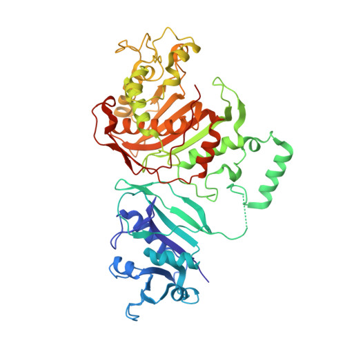
Nonconserved residues Ala287 and Ser290 of the Cryptosporidium hominis thymidylate synthase domain facilitate its rapid rate of catalysis

Doan, L.T.Martucci, W.E.Vargo, M.A.Atreya, C.E.Anderson, K.S.

(2007) Biochemistry 46: 8379-8391

  • DOI: https://doi.org/10.1021/bi700531r
  • Primary Citation of Related Structures:  
    2OIP

  • PubMed Abstract: 

    Cryptosporidium hominis TS-DHFR exhibits an unusually high rate of catalysis at the TS domain, at least 10-fold greater than those of other TS enzymes. Using site-directed mutagenesis, we have mutated residues Ala287 and Ser290 in the folate-binding helix to phenylalanine and glycine, respectively, the corresponding residues in human and most other TS enzymes. Our results show that the mutant A287F, the mutant S290G, and the double mutant all have reduced affinities for methylene tetrahydrofolate and reduced rates of reaction at the TS domain. Interestingly, the S290G mutant enzyme had the lowest TS activity, with a catalytic efficiency approximately 200-fold lower than that of the wild type (WT). The rate of conformational change of the S290G mutant is approximately 80 times slower than that of WT, resulting in a change in the rate-limiting step from hydride transfer to covalent ternary complex formation. We have determined the crystal structure of ligand-bound S290G mutant enzyme, which shows that the primary effect of the mutation is an increase in the distance between the TS ligands. The kinetic and crystal structure data presented here provide the first evidence explaining the unusually fast TS rate in C. hominis.


  • Organizational Affiliation

    Department of Pharmacology, Yale University School of Medicine, 333 Cedar Street, New Haven, Connecticut 06520, USA.


Macromolecules
Find similar proteins by:  (by identity cutoff)  |  3D Structure
Entity ID: 1
MoleculeChains Sequence LengthOrganismDetailsImage
Chain A, crystal structure of Dhfr
A, B, C, D, E
519Cryptosporidium hominisMutation(s): 1 
EC: 2.1.1.45 (PDB Primary Data), 1.5.1.3 (PDB Primary Data)
Entity Groups  
Sequence Clusters30% Identity50% Identity70% Identity90% Identity95% Identity100% Identity
Sequence Annotations
Expand
  • Reference Sequence
Small Molecules
Ligands 4 Unique
IDChains Name / Formula / InChI Key2D Diagram3D Interactions
NDP
Query on NDP

Download Ideal Coordinates CCD File 
I [auth A],
M [auth B],
Q [auth C],
U [auth D],
Y [auth E]
NADPH DIHYDRO-NICOTINAMIDE-ADENINE-DINUCLEOTIDE PHOSPHATE
C21 H30 N7 O17 P3
ACFIXJIJDZMPPO-NNYOXOHSSA-N
CB3
Query on CB3

Download Ideal Coordinates CCD File 
G [auth A],
K [auth B],
O [auth C],
S [auth D],
W [auth E]
10-PROPARGYL-5,8-DIDEAZAFOLIC ACID
C24 H23 N5 O6
LTKHPMDRMUCUEB-IBGZPJMESA-N
MTX
Query on MTX

Download Ideal Coordinates CCD File 
H [auth A],
L [auth B],
P [auth C],
T [auth D],
X [auth E]
METHOTREXATE
C20 H22 N8 O5
FBOZXECLQNJBKD-ZDUSSCGKSA-N
UMP
Query on UMP

Download Ideal Coordinates CCD File 
F [auth A],
J [auth B],
N [auth C],
R [auth D],
V [auth E]
2'-DEOXYURIDINE 5'-MONOPHOSPHATE
C9 H13 N2 O8 P
JSRLJPSBLDHEIO-SHYZEUOFSA-N
Experimental Data & Validation

Experimental Data

  • Method: X-RAY DIFFRACTION
  • Resolution: 2.80 Å
  • R-Value Free: 0.259 
  • R-Value Work: 0.222 
  • R-Value Observed: 0.222 
  • Space Group: C 1 2 1
Unit Cell:
Length ( Å )Angle ( ˚ )
a = 215.026α = 90
b = 116.204β = 94.27
c = 216.601γ = 90
Software Package:
Software NamePurpose
DENZOdata reduction
SCALEPACKdata scaling
SOLOMONphasing
CNSrefinement
PDB_EXTRACTdata extraction
CBASSdata collection
HKL-2000data reduction
AMoREphasing

Structure Validation

View Full Validation Report


Ligand Structure Quality Assessment 


Entry History 

Deposition Data

Revision History  (Full details and data files)

  • Version 1.0: 2007-07-17
    Type: Initial release
  • Version 1.1: 2008-05-01
    Changes: Version format compliance
  • Version 1.2: 2011-07-13
    Changes: Version format compliance
  • Version 1.3: 2017-10-18
    Changes: Refinement description
  • Version 1.4: 2021-10-20
    Changes: Database references, Derived calculations
  • Version 1.5: 2023-08-30
    Changes: Data collection, Refinement description