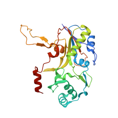
2PHN

Crystal structure of an amide bond forming F420-gamma glutamyl ligase from Archaeoglobus fulgidus


Experimental Data Snapshot

  • Method: X-RAY DIFFRACTION
  • Resolution: 1.35 Å
  • R-Value Free: 0.190 
  • R-Value Work: 0.160 
  • R-Value Observed: 0.162 

wwPDB Validation   3D Report Full Report


Ligand Structure Quality Assessment 


This is version 1.5 of the entry. See complete history


Literature

Structure of an Amide Bond Forming F(420):gammagamma-glutamyl Ligase from Archaeoglobus Fulgidus - A Member of a New Family of Non-ribosomal Peptide Synthases.

Nocek, B.Evdokimova, E.Proudfoot, M.Kudritska, M.Grochowski, L.L.White, R.H.Savchenko, A.Yakunin, A.F.Edwards, A.Joachimiak, A.

(2007) J Mol Biol 372: 456-469

  • DOI: https://doi.org/10.1016/j.jmb.2007.06.063
  • Primary Citation of Related Structures:  
    2G9I, 2PHN

  • PubMed Abstract: 

    F(420) is a flavin-like redox-active coenzyme commonly used by archaea and some eubacteria in a variety of biochemical reactions in methanogenesis, the formation of secondary metabolites, the degradation of nitroaromatic compounds, activation of nitroimidazofurans, and F(420)-dependent photolysis in DNA repair. Coenzyme F(420)-2 biosynthesis from 7,8-didemethyl-8-hydroxy-5-deazariboflavin (Fo) and lactaldehyde involves six enzymatic steps and five proteins (CofA, CofB, CofC, CofD, and CofE). CofE, a F(420)-0:gamma-glutamyl ligase, is responsible for the last two enzymatic steps; it catalyses the GTP-dependent addition of two L-glutamate residues to F(420)-0 to form F(420)-2. CofE is found in archaea, the aerobic actinomycetes, and cyanobacteria. Here, we report the first crystal structure of the apo-F(420)-0:gamma-glutamyl ligase (CofE-AF) from Archaeoglobus fulgidus and its complex with GDP at 2.5 A and 1.35 A resolution, respectively. The structure of CofE-AF reveals a novel protein fold with an intertwined, butterfly-like dimer formed by two-domain monomers. GDP and Mn(2+) are bound within the putative active site in a large groove at the dimer interface. We show that the enzyme adds a glutamate residue to both F(420)-0 and F(420)-1 in two distinct steps. CofE represents the first member of a new structural family of non-ribosomal peptide synthases.


  • Organizational Affiliation

    Midwest Center for Structural Genomics and Structural Biology Center, Biosciences, Argonne National Laboratory, Argonne, IL 60439, USA.


Macromolecules
Find similar proteins by:  (by identity cutoff)  |  3D Structure
Entity ID: 1
MoleculeChains Sequence LengthOrganismDetailsImage
F420-0:gamma-glutamyl ligase
A, B
254Archaeoglobus fulgidus DSM 4304Mutation(s): 0 
Gene Names: cofEAF_2256
EC: 6.3.2
UniProt
Find proteins for O28028 (Archaeoglobus fulgidus (strain ATCC 49558 / DSM 4304 / JCM 9628 / NBRC 100126 / VC-16))
Explore O28028 
Go to UniProtKB:  O28028
Entity Groups  
Sequence Clusters30% Identity50% Identity70% Identity90% Identity95% Identity100% Identity
UniProt GroupO28028
Sequence Annotations
Expand
  • Reference Sequence
Small Molecules
Ligands 5 Unique
IDChains Name / Formula / InChI Key2D Diagram3D Interactions
GDP
Query on GDP

Download Ideal Coordinates CCD File 
H [auth A],
Q [auth B]
GUANOSINE-5'-DIPHOSPHATE
C10 H15 N5 O11 P2
QGWNDRXFNXRZMB-UUOKFMHZSA-N
GOL
Query on GOL

Download Ideal Coordinates CCD File 
M [auth A]GLYCEROL
C3 H8 O3
PEDCQBHIVMGVHV-UHFFFAOYSA-N
EDO
Query on EDO

Download Ideal Coordinates CCD File 
I [auth A],
J [auth A],
K [auth A],
L [auth A]
1,2-ETHANEDIOL
C2 H6 O2
LYCAIKOWRPUZTN-UHFFFAOYSA-N
ACT
Query on ACT

Download Ideal Coordinates CCD File 
E [auth A],
F [auth A],
G [auth A],
P [auth B]
ACETATE ION
C2 H3 O2
QTBSBXVTEAMEQO-UHFFFAOYSA-M
MN
Query on MN

Download Ideal Coordinates CCD File 
C [auth A],
D [auth A],
N [auth B],
O [auth B]
MANGANESE (II) ION
Mn
WAEMQWOKJMHJLA-UHFFFAOYSA-N
Experimental Data & Validation

Experimental Data

  • Method: X-RAY DIFFRACTION
  • Resolution: 1.35 Å
  • R-Value Free: 0.190 
  • R-Value Work: 0.160 
  • R-Value Observed: 0.162 
  • Space Group: P 41 21 2
Unit Cell:
Length ( Å )Angle ( ˚ )
a = 98.367α = 90
b = 98.367β = 90
c = 94.347γ = 90
Software Package:
Software NamePurpose
REFMACrefinement
SBC-Collectdata collection
HKL-3000data reduction
HKL-3000data scaling
MOLREPphasing

Structure Validation

View Full Validation Report


Ligand Structure Quality Assessment 


Entry History 

Revision History  (Full details and data files)

  • Version 1.0: 2007-05-15
    Type: Initial release
  • Version 1.1: 2008-05-01
    Changes: Version format compliance
  • Version 1.2: 2011-07-13
    Changes: Non-polymer description, Version format compliance
  • Version 1.3: 2011-10-19
    Changes: Derived calculations
  • Version 1.4: 2023-08-30
    Changes: Data collection, Database references, Derived calculations, Refinement description
  • Version 1.5: 2023-12-27
    Changes: Derived calculations