3Q5E | pdb_00003q5e

crystal structure of human Atlastin-1 (residues 1-447) bound to GDP, crystal form 2


Experimental Data Snapshot

  • Method: X-RAY DIFFRACTION
  • Resolution: 3.01 Å
  • R-Value Free: 
    0.273 (Depositor), 0.270 (DCC) 
  • R-Value Work: 
    0.197 (Depositor), 0.190 (DCC) 
  • R-Value Observed: 
    0.200 (Depositor) 

wwPDB Validation   3D Report Full Report


Ligand Structure Quality Assessment 


This is version 1.2 of the entry. See complete history


Literature

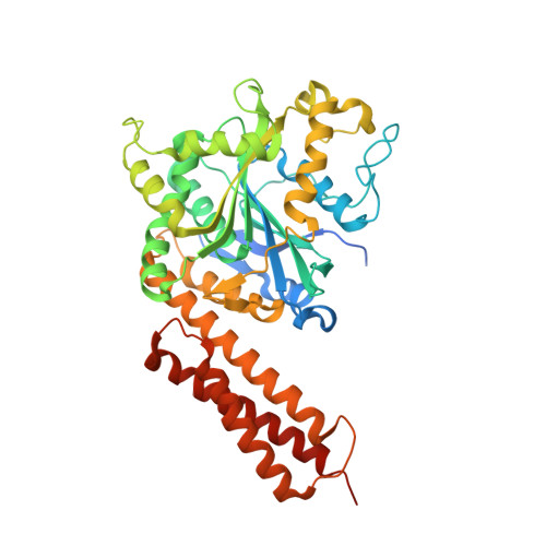
Structural basis for the nucleotide-dependent dimerization of the large G protein atlastin-1/SPG3A.

Byrnes, L.J.Sondermann, H.

(2011) Proc Natl Acad Sci U S A 108: 2216-2221

  • DOI: https://doi.org/10.1073/pnas.1012792108
  • Primary Citation of Related Structures:  
    3Q5D, 3Q5E

  • PubMed Abstract: 

    The large GTPase atlastin belongs to the dynamin superfamily that has been widely implicated in facilitating membrane tubulation, fission, and in select cases, fusion. Mutations spread across atlastin isoform 1 (atlastin-1) have been identified in patients suffering from hereditary spastic paraplegia (HSP), a neurodegenerative disorder affecting motor neuron function in the lower extremities. On a molecular level, atlastin-1 associates with high membrane curvature and fusion events at the endoplasmic reticulum and cis-Golgi. Here we report crystal structures of atlastin-1 comprising the G and middle domains in two different conformations. Although the orientation of the middle domain relative to the G domain is different in the two structures, both reveal dimeric assemblies with a common, GDP-bound G domain dimer. In contrast, dimer formation in solution is observed only in the presence of GTP and transition state analogs, similar to other G proteins that are activated by nucleotide-dependent dimerization. Analyses of solution scattering data suggest that upon nucleotide binding, the protein adopts a somewhat extended, dimeric conformation that is reminiscent of one of the two crystal structures. These structural studies suggest a model for nucleotide-dependent regulation of atlastin with implications for membrane fusion. This mechanism is affected in several mutants associated with HSP, providing insights into disease pathogenesis.


  • Organizational Affiliation
    • Department of Molecular Medicine, College of Veterinary Medicine, Cornell University, Ithaca, NY 14853, USA.

Macromolecules
Find similar proteins by:  (by identity cutoff)  |  3D Structure
Entity ID: 1
MoleculeChains Sequence LengthOrganismDetailsImage
Atlastin-1A,
B [auth C],
C [auth E],
D [auth G]
447Homo sapiensMutation(s): 0 
Gene Names: ATL1GBP3SPG3A
EC: 3.6.5
UniProt & NIH Common Fund Data Resources
Find proteins for Q8WXF7 (Homo sapiens)
Explore Q8WXF7 
Go to UniProtKB:  Q8WXF7
PHAROS:  Q8WXF7
GTEx:  ENSG00000198513 
Entity Groups  
Sequence Clusters30% Identity50% Identity70% Identity90% Identity95% Identity100% Identity
UniProt GroupQ8WXF7
Sequence Annotations
Expand
  • Reference Sequence
Small Molecules
Ligands 2 Unique
IDChains Name / Formula / InChI Key2D Diagram3D Interactions
GDP
Query on GDP

Download Ideal Coordinates CCD File 
E [auth A],
G [auth C],
I [auth E],
K [auth G]
GUANOSINE-5'-DIPHOSPHATE
C10 H15 N5 O11 P2
QGWNDRXFNXRZMB-UUOKFMHZSA-N
MG
Query on MG

Download Ideal Coordinates CCD File 
F [auth A],
H [auth C],
J [auth E],
L [auth G]
MAGNESIUM ION
Mg
JLVVSXFLKOJNIY-UHFFFAOYSA-N
Experimental Data & Validation

Experimental Data

  • Method: X-RAY DIFFRACTION
  • Resolution: 3.01 Å
  • R-Value Free:  0.273 (Depositor), 0.270 (DCC) 
  • R-Value Work:  0.197 (Depositor), 0.190 (DCC) 
  • R-Value Observed: 0.200 (Depositor) 
Space Group: P 21 21 21
Unit Cell:
Length ( Å )Angle ( ˚ )
a = 104.394α = 90
b = 133.549β = 90
c = 175.835γ = 90
Software Package:
Software NamePurpose
ADSCdata collection
PHENIXmodel building
PHASERphasing
PHENIXrefinement
HKL-2000data reduction
HKL-2000data scaling
PHENIXphasing

Structure Validation

View Full Validation Report


Ligand Structure Quality Assessment 


Entry History 

Deposition Data

Revision History  (Full details and data files)

  • Version 1.0: 2011-01-19
    Type: Initial release
  • Version 1.1: 2011-07-13
    Changes: Version format compliance
  • Version 1.2: 2024-02-21
    Changes: Data collection, Database references, Derived calculations