4CHX

Crystal structure of MltC in complex with disaccharide pentapeptide DHl89


Experimental Data Snapshot

  • Method: X-RAY DIFFRACTION
  • Resolution: 2.45 Å
  • R-Value Free: 0.248 
  • R-Value Work: 0.192 
  • R-Value Observed: 0.196 

wwPDB Validation   3D Report Full Report


Ligand Structure Quality Assessment 


This is version 3.1 of the entry. See complete history


Literature

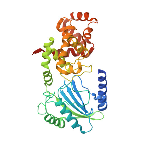
Structure and Cell Wall Cleavage by Modular Lytic Transglycosylase Mltc of Escherichia Coli.

Artola-Recolons, C.Lee, M.Bernardo-Garcia, N.Blazquez, B.Hesek, D.Bartual, S.G.Mahasenan, K.V.Lastochkin, E.Pi, H.Boggess, B.Meindl, K.Uson, I.Fisher, J.F.Mobashery, S.Hermoso, J.A.

(2014) ACS Chem Biol 9: 2058

  • DOI: https://doi.org/10.1021/cb500439c
  • Primary Citation of Related Structures:  
    4C5F, 4CFO, 4CFP, 4CHX

  • PubMed Abstract: 

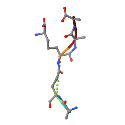
    The lytic transglycosylases are essential bacterial enzymes that catalyze the nonhydrolytic cleavage of the glycan strands of the bacterial cell wall. We describe here the structural and catalytic properties of MltC, one of the seven lytic transglycosylases found in the genome of the Gram-negative bacterium Escherichia coli. The 2.3 Å resolution X-ray structure of a soluble construct of MltC shows a unique, compared to known lytic transglycosylase structures, two-domain structure characterized by an expansive active site of 53 Å length extending through an interface between the domains. The structures of three complexes of MltC with cell wall analogues suggest the positioning of the peptidoglycan in the active site both as a substrate and as a product. One complex is suggested to correspond to an intermediate in the course of sequential and exolytic cleavage of the peptidoglycan. Moreover, MltC partitioned its reactive oxocarbenium-like intermediate between trapping by the C6-hydroxyl of the muramyl moiety (lytic transglycosylase activity, the major path) and by water (muramidase activity). Genomic analysis identifies the presence of an MltC homologue in no less than 791 bacterial genomes. While the role of MltC in cell wall assembly and maturation remains uncertain, we propose a functional role for this enzyme as befits the uniqueness of its two-domain structure.


  • Organizational Affiliation

    Department of Crystallography and Structural Biology, Inst. Química-Física "Rocasolano", CSIC , Serrano 119, 28006 Madrid, Spain.


Macromolecules
Find similar proteins by:  (by identity cutoff)  |  3D Structure
Entity ID: 1
MoleculeChains Sequence LengthOrganismDetailsImage
Membrane-bound lytic murein transglycosylase C
A, B
341Escherichia coliMutation(s): 0 
Gene Names: mltCyggZb2963JW5481
EC: 4.2.2
UniProt
Find proteins for P0C066 (Escherichia coli (strain K12))
Explore P0C066 
Go to UniProtKB:  P0C066
Entity Groups  
Sequence Clusters30% Identity50% Identity70% Identity90% Identity95% Identity100% Identity
UniProt GroupP0C066
Sequence Annotations
Expand
  • Reference Sequence

Find similar proteins by:  Sequence   |   3D Structure  

Entity ID: 2
MoleculeChains Sequence LengthOrganismDetailsImage
NAG-ANHMUR-PENTAPEPTIDE5synthetic constructMutation(s): 0 
Sequence Annotations
Expand
  • Reference Sequence
Small Molecules
Ligands 2 Unique
IDChains Name / Formula / InChI Key2D Diagram3D Interactions
AH0
Query on AH0

Download Ideal Coordinates CCD File 
E [auth C]2-(2-ACETYLAMINO-4-HYDROXY-6,8-DIOXA-BICYCLO[3.2.1]OCT-3-YLOXY)-PROPIONIC ACID
C11 H17 N O7
ZFEGYUMHFZOYIY-YVNCZSHWSA-N
NAG
Query on NAG

Download Ideal Coordinates CCD File 
D [auth A]2-acetamido-2-deoxy-beta-D-glucopyranose
C8 H15 N O6
OVRNDRQMDRJTHS-FMDGEEDCSA-N
Modified Residues  1 Unique
IDChains TypeFormula2D DiagramParent
API
Query on API
C
L-PEPTIDE LINKINGC7 H14 N2 O4LYS
Experimental Data & Validation

Experimental Data

  • Method: X-RAY DIFFRACTION
  • Resolution: 2.45 Å
  • R-Value Free: 0.248 
  • R-Value Work: 0.192 
  • R-Value Observed: 0.196 
  • Space Group: P 1 21 1
Unit Cell:
Length ( Å )Angle ( ˚ )
a = 50.16α = 90
b = 115β = 93.21
c = 61.28γ = 90
Software Package:
Software NamePurpose
PHENIXmodel building
SCALAdata scaling
iMOSFLMphasing
SCALAphasing
MOLREPphasing
PHENIXphasing
PHENIXrefinement

Structure Validation

View Full Validation Report


Ligand Structure Quality Assessment 


Entry History 

Deposition Data

Revision History  (Full details and data files)

  • Version 1.0: 2014-07-23
    Type: Initial release
  • Version 1.1: 2014-10-01
    Changes: Database references
  • Version 1.2: 2020-07-29
    Type: Remediation
    Reason: Carbohydrate remediation
    Changes: Advisory, Data collection, Derived calculations, Other, Structure summary
  • Version 2.0: 2022-12-07
    Changes: Advisory, Atomic model, Data collection, Database references, Derived calculations, Polymer sequence, Source and taxonomy, Structure summary
  • Version 3.0: 2023-11-15
    Changes: Atomic model, Data collection, Derived calculations
  • Version 3.1: 2024-01-31
    Changes: Refinement description