4XIC

ANTPHD WITH 15BP di-thioate modified DNA DUPLEX


Experimental Data Snapshot

  • Method: X-RAY DIFFRACTION
  • Resolution: 2.69 Å
  • R-Value Free: 0.272 
  • R-Value Work: 0.216 
  • R-Value Observed: 0.219 

wwPDB Validation   3D Report Full Report


This is version 1.3 of the entry. See complete history


Literature

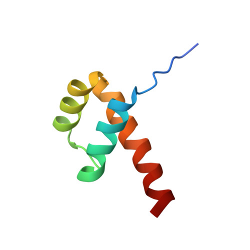
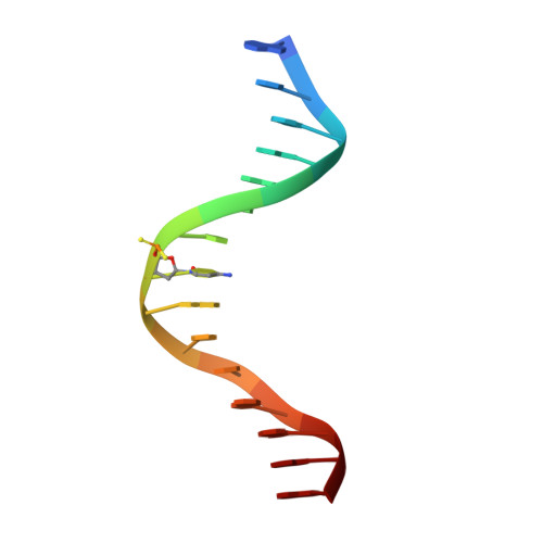
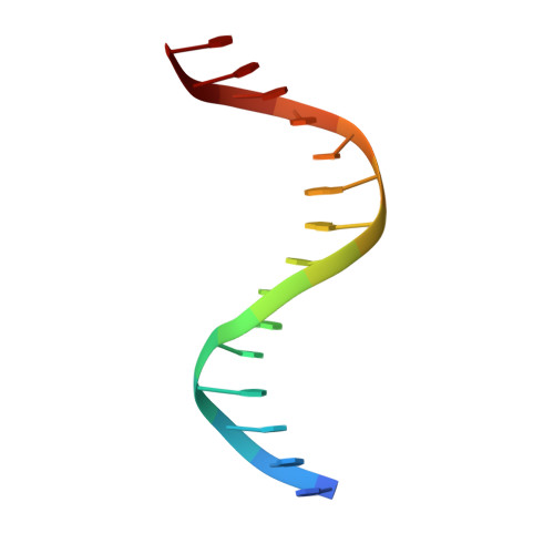
Entropic Enhancement of Protein-DNA Affinity by Oxygen-to-Sulfur Substitution in DNA Phosphate.

Zandarashvili, L.Nguyen, D.Anderson, K.M.White, M.A.Gorenstein, D.G.Iwahara, J.

(2015) Biophys J 109: 1026-1037

  • DOI: https://doi.org/10.1016/j.bpj.2015.07.032
  • Primary Citation of Related Structures:  
    4XIC, 4XID

  • PubMed Abstract: 

    Dithioation of DNA phosphate is known to enhance binding affinities, at least for some proteins. We mechanistically characterized this phenomenon for the Antennapedia homeodomain-DNA complex by integrated use of fluorescence, isothermal titration calorimetry, NMR spectroscopy, and x-ray crystallography. By fluorescence and isothermal titration calorimetry, we found that this affinity enhancement is entropy driven. By NMR, we investigated the ionic hydrogen bonds and internal motions of lysine side-chain NH3(+) groups involved in ion pairs with DNA. By x-ray crystallography, we compared the structures of the complexes with and without dithioation of the phosphate. Our NMR and x-ray data show that the lysine side chain in contact with the DNA phosphate becomes more dynamic upon dithioation. Our thermodynamic, structural, and dynamic investigations collectively suggest that the affinity enhancement by the oxygen-to-sulfur substitution in DNA phosphate is largely due to an entropic gain arising from mobilization of the intermolecular ion pair at the protein-DNA interface.


  • Organizational Affiliation

    Department of Biochemistry and Molecular Biology, Sealy Center for Structural Biology and Molecular Biophysics, University of Texas Medical Branch, Galveston, Texas.


Macromolecules

Find similar proteins by:  (by identity cutoff)  |  3D Structure
Entity ID: 1
MoleculeChains Sequence LengthOrganismDetailsImage
Homeotic protein antennapedia
A, D
61Drosophila melanogasterMutation(s): 1 
Gene Names: AntpCG1028
UniProt
Find proteins for P02833 (Drosophila melanogaster)
Explore P02833 
Go to UniProtKB:  P02833
Entity Groups  
Sequence Clusters30% Identity50% Identity70% Identity90% Identity95% Identity100% Identity
UniProt GroupP02833
Sequence Annotations
Expand
  • Reference Sequence

Find similar nucleic acids by:  Sequence   |   3D Structure  

Entity ID: 2
MoleculeChains LengthOrganismImage
DNA (5'-D(*AP*GP*AP*AP*AP*GP*CP*(C2S)P*AP*TP*TP*AP*GP*AP*G)-3')
B, E
15unidentified
Sequence Annotations
Expand
  • Reference Sequence

Find similar nucleic acids by:  Sequence   |   3D Structure  

Entity ID: 3
MoleculeChains LengthOrganismImage
DNA (5'-D(*TP*CP*TP*CP*TP*AP*AP*TP*GP*GP*CP*TP*TP*TP*C)-3')
C, F
15unidentified
Sequence Annotations
Expand
  • Reference Sequence
Experimental Data & Validation

Experimental Data

  • Method: X-RAY DIFFRACTION
  • Resolution: 2.69 Å
  • R-Value Free: 0.272 
  • R-Value Work: 0.216 
  • R-Value Observed: 0.219 
  • Space Group: P 43 21 2
Unit Cell:
Length ( Å )Angle ( ˚ )
a = 96.539α = 90
b = 96.539β = 90
c = 89.546γ = 90
Software Package:
Software NamePurpose
PHENIXrefinement
HKL-2000data scaling
PDB_EXTRACTdata extraction
Cootmodel building
HKL-2000data reduction

Structure Validation

View Full Validation Report


Entry History 

Deposition Data

Revision History  (Full details and data files)

  • Version 1.0: 2015-11-25
    Type: Initial release
  • Version 1.1: 2016-03-16
    Changes: Data collection
  • Version 1.2: 2017-11-22
    Changes: Derived calculations, Refinement description
  • Version 1.3: 2023-09-27
    Changes: Data collection, Database references, Derived calculations, Refinement description