5H3D

Helical structure of membrane tubules decorated by ACAP1 (BARPH doamin) protein by cryo-electron microscopy and MD simulation


Experimental Data Snapshot

  • Method: ELECTRON MICROSCOPY
  • Resolution: 14.0 Å
  • Aggregation State: HELICAL ARRAY 
  • Reconstruction Method: HELICAL 

wwPDB Validation   3D Report Full Report


This is version 1.3 of the entry. See complete history

Re-refinement Note

This entry reflects an alternative modeling of the original data in: 4CKG


Literature

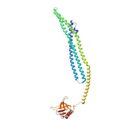
ACAP1 assembles into an unusual protein lattice for membrane deformation through multiple stages.

Chan, C.Pang, X.Zhang, Y.Niu, T.Yang, S.Zhao, D.Li, J.Lu, L.Hsu, V.W.Zhou, J.Sun, F.Fan, J.

(2019) PLoS Comput Biol 15: e1007081-e1007081

  • DOI: https://doi.org/10.1371/journal.pcbi.1007081
  • Primary Citation of Related Structures:  
    5H3D

  • PubMed Abstract: 

    Studies on the Bin-Amphiphysin-Rvs (BAR) domain have advanced a fundamental understanding of how proteins deform membrane. We previously showed that a BAR domain in tandem with a Pleckstrin Homology (PH domain) underlies the assembly of ACAP1 (Arfgap with Coil-coil, Ankryin repeat, and PH domain I) into an unusual lattice structure that also uncovers a new paradigm for how a BAR protein deforms membrane. Here, we initially pursued computation-based refinement of the ACAP1 lattice to identify its critical protein contacts. Simulation studies then revealed how ACAP1, which dimerizes into a symmetrical structure in solution, is recruited asymmetrically to the membrane through dynamic behavior. We also pursued electron microscopy (EM)-based structural studies, which shed further insight into the dynamic nature of the ACAP1 lattice assembly. As ACAP1 is an unconventional BAR protein, our findings broaden the understanding of the mechanistic spectrum by which proteins assemble into higher-ordered structures to achieve membrane deformation.


  • Organizational Affiliation

    Department of Materials Science and Engineering, City University of Hong Kong, Hong Kong, China.


Macromolecules
Find similar proteins by:  (by identity cutoff)  |  3D Structure
Entity ID: 1
MoleculeChains Sequence LengthOrganismDetailsImage
Arf-GAP with coiled-coil, ANK repeat and PH domain-containing protein 1
A, B, C, D
382Homo sapiensMutation(s): 0 
Gene Names: ACAP1CENTB1KIAA0050
UniProt & NIH Common Fund Data Resources
Find proteins for Q15027 (Homo sapiens)
Explore Q15027 
Go to UniProtKB:  Q15027
PHAROS:  Q15027
GTEx:  ENSG00000072818 
Entity Groups  
Sequence Clusters30% Identity50% Identity70% Identity90% Identity95% Identity100% Identity
UniProt GroupQ15027
Sequence Annotations
Expand
  • Reference Sequence
Experimental Data & Validation

Experimental Data

  • Method: ELECTRON MICROSCOPY
  • Resolution: 14.0 Å
  • Aggregation State: HELICAL ARRAY 
  • Reconstruction Method: HELICAL 

Structure Validation

View Full Validation Report


Entry History & Funding Information

Deposition Data


Funding OrganizationLocationGrant Number
National Natural Science Foundation of ChinaChina--

Revision History  (Full details and data files)

  • Version 1.0: 2019-01-16
    Type: Initial release
  • Version 1.1: 2019-11-06
    Changes: Data collection, Other
  • Version 1.2: 2020-02-12
    Changes: Database references
  • Version 1.3: 2024-03-20
    Changes: Data collection, Database references, Derived calculations