5NJH

Triazolopyrimidines stabilize microtubules by binding to the vinca inhibitor site of tubulin


Experimental Data Snapshot

  • Method: X-RAY DIFFRACTION
  • Resolution: 2.39 Å
  • R-Value Free: 0.237 
  • R-Value Work: 0.217 
  • R-Value Observed: 0.218 

Starting Model: experimental
View more details

wwPDB Validation   3D Report Full Report


Ligand Structure Quality Assessment 


This is version 1.2 of the entry. See complete history


Literature

Triazolopyrimidines Are Microtubule-Stabilizing Agents that Bind the Vinca Inhibitor Site of Tubulin.

Saez-Calvo, G.Sharma, A.Balaguer, F.A.Barasoain, I.Rodriguez-Salarichs, J.Olieric, N.Munoz-Hernandez, H.Berbis, M.A.Wendeborn, S.Penalva, M.A.Matesanz, R.Canales, A.Prota, A.E.Jimenez-Barbero, J.Andreu, J.M.Lamberth, C.Steinmetz, M.O.Diaz, J.F.

(2017) Cell Chem Biol 24: 737-750.e6

  • DOI: https://doi.org/10.1016/j.chembiol.2017.05.016
  • Primary Citation of Related Structures:  
    5NJH

  • PubMed Abstract: 

    Microtubule-targeting agents (MTAs) are some of the clinically most successful anti-cancer drugs. Unfortunately, instances of multidrug resistances to MTA have been reported, which highlights the need for developing MTAs with different mechanistic properties. One less explored class of MTAs are [1,2,4]triazolo[1,5-a]pyrimidines (TPs). These cytotoxic compounds are microtubule-stabilizing agents that inexplicably bind to vinblastine binding site on tubulin, which is typically targeted by microtubule-destabilizing agents. Here we used cellular, biochemical, and structural biology approaches to address this apparent discrepancy. Our results establish TPs as vinca-site microtubule-stabilizing agents that promote longitudinal tubulin contacts in microtubules, in contrast to classical microtubule-stabilizing agents that primarily promote lateral contacts. Additionally we observe that TPs studied here are not affected by p-glycoprotein overexpression, and suggest that TPs are promising ligands against multidrug-resistant cancer cells.


  • Organizational Affiliation

    Department of Physical and Chemical Biology, Centro de Investigaciones Biológicas, Consejo Superior de Investigaciones Científicas, Ramiro de Maeztu 9, 28040 Madrid, Spain.


Macromolecules
Find similar proteins by:  (by identity cutoff)  |  3D Structure
Entity ID: 1
MoleculeChains Sequence LengthOrganismDetailsImage
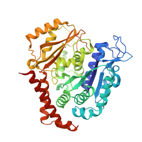
Tubulin alpha-1B chain
A, C
450Bos taurusMutation(s): 0 
UniProt
Find proteins for P81947 (Bos taurus)
Explore P81947 
Go to UniProtKB:  P81947
Entity Groups  
Sequence Clusters30% Identity50% Identity70% Identity90% Identity95% Identity100% Identity
UniProt GroupP81947
Sequence Annotations
Expand
  • Reference Sequence
Find similar proteins by:  (by identity cutoff)  |  3D Structure
Entity ID: 2
MoleculeChains Sequence LengthOrganismDetailsImage
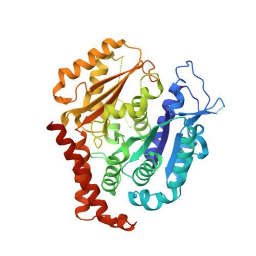
Tubulin beta-2B chain
B, D
445Bos taurusMutation(s): 0 
UniProt
Find proteins for Q6B856 (Bos taurus)
Explore Q6B856 
Go to UniProtKB:  Q6B856
Entity Groups  
Sequence Clusters30% Identity50% Identity70% Identity90% Identity95% Identity100% Identity
UniProt GroupQ6B856
Sequence Annotations
Expand
  • Reference Sequence
Find similar proteins by:  (by identity cutoff)  |  3D Structure
Entity ID: 3
MoleculeChains Sequence LengthOrganismDetailsImage
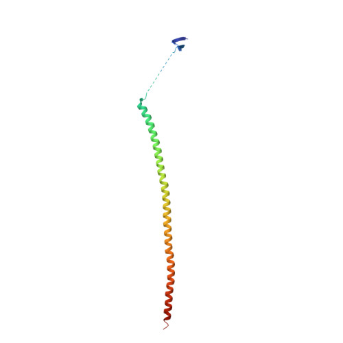
Stathmin-4143Rattus norvegicusMutation(s): 0 
Gene Names: Stmn4
UniProt
Find proteins for P63043 (Rattus norvegicus)
Explore P63043 
Go to UniProtKB:  P63043
Entity Groups  
Sequence Clusters30% Identity50% Identity70% Identity90% Identity95% Identity100% Identity
UniProt GroupP63043
Sequence Annotations
Expand
  • Reference Sequence
Find similar proteins by:  (by identity cutoff)  |  3D Structure
Entity ID: 4
MoleculeChains Sequence LengthOrganismDetailsImage
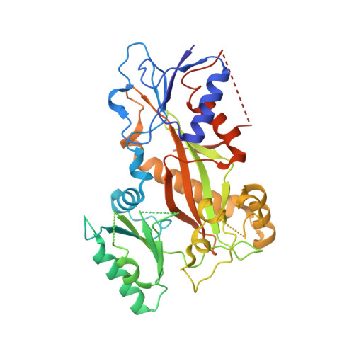
tubulin tyrosine ligase TTL384Gallus gallusMutation(s): 0 
Gene Names: TTL
Entity Groups  
Sequence Clusters30% Identity50% Identity70% Identity90% Identity95% Identity100% Identity
Sequence Annotations
Expand
  • Reference Sequence
Small Molecules
Ligands 5 Unique
IDChains Name / Formula / InChI Key2D Diagram3D Interactions
GTP
Query on GTP

Download Ideal Coordinates CCD File 
G [auth A],
L [auth C]
GUANOSINE-5'-TRIPHOSPHATE
C10 H16 N5 O14 P3
XKMLYUALXHKNFT-UUOKFMHZSA-N
ACP
Query on ACP

Download Ideal Coordinates CCD File 
Q [auth F]PHOSPHOMETHYLPHOSPHONIC ACID ADENYLATE ESTER
C11 H18 N5 O12 P3
UFZTZBNSLXELAL-IOSLPCCCSA-N
GDP
Query on GDP

Download Ideal Coordinates CCD File 
I [auth B],
N [auth D]
GUANOSINE-5'-DIPHOSPHATE
C10 H15 N5 O11 P2
QGWNDRXFNXRZMB-UUOKFMHZSA-N
8Z8
Query on 8Z8

Download Ideal Coordinates CCD File 
K [auth B]5-chloranyl-7-[(1~{R},5~{S})-3-methoxy-8-azabicyclo[3.2.1]octan-8-yl]-6-[2,4,6-tris(fluoranyl)phenyl]-[1,2,4]triazolo[1,5-a]pyrimidine
C19 H17 Cl F3 N5 O
QLFREGOMFNYDON-GDNZZTSVSA-N
MG
Query on MG

Download Ideal Coordinates CCD File 
H [auth A],
J [auth B],
M [auth C],
O [auth D],
P [auth F]
MAGNESIUM ION
Mg
JLVVSXFLKOJNIY-UHFFFAOYSA-N
Experimental Data & Validation

Experimental Data

  • Method: X-RAY DIFFRACTION
  • Resolution: 2.39 Å
  • R-Value Free: 0.237 
  • R-Value Work: 0.217 
  • R-Value Observed: 0.218 
  • Space Group: P 21 21 21
Unit Cell:
Length ( Å )Angle ( ˚ )
a = 103.43α = 90
b = 158.353β = 90
c = 173.458γ = 90
Software Package:
Software NamePurpose
PHENIXrefinement
XDSdata reduction
XSCALEdata scaling
PHENIXphasing

Structure Validation

View Full Validation Report


Ligand Structure Quality Assessment 


Entry History 

Deposition Data

Revision History  (Full details and data files)

  • Version 1.0: 2017-06-21
    Type: Initial release
  • Version 1.1: 2017-07-05
    Changes: Database references
  • Version 1.2: 2024-01-17
    Changes: Advisory, Data collection, Database references, Derived calculations, Refinement description