6MZG

Structural Basis of Tubulin Recruitment and Assembly by Microtubule Polymerases with Tumor Overexpressed Gene (TOG) Domain Arrays


Experimental Data Snapshot

  • Method: X-RAY DIFFRACTION
  • Resolution: 3.21 Å
  • R-Value Free: 0.234 
  • R-Value Work: 0.175 
  • R-Value Observed: 0.180 

wwPDB Validation   3D Report Full Report


Ligand Structure Quality Assessment 


This is version 1.1 of the entry. See complete history


Literature

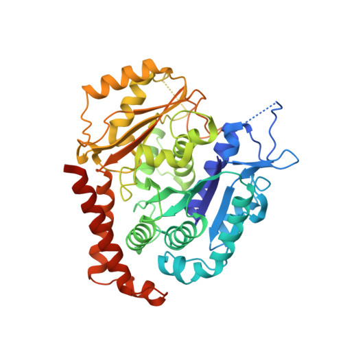
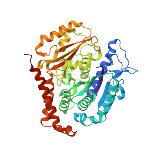
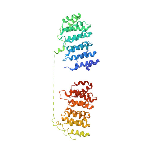
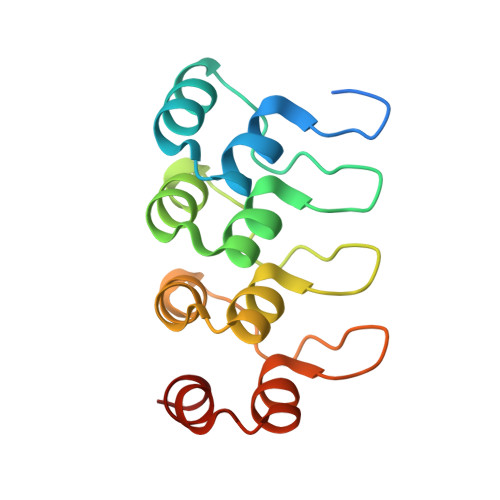
Structural basis of tubulin recruitment and assembly by microtubule polymerases with Tumor Overexpressed Gene (TOG) domain arrays.

Nithianantham, S.Cook, B.D.Beans, M.Guo, F.Chang, F.Al-Bassam, J.

(2018) Elife 7

  • DOI: https://doi.org/10.7554/eLife.38922
  • Primary Citation of Related Structures:  
    6MZE, 6MZF, 6MZG

  • PubMed Abstract: 
  • XMAP215/Stu2/Alp14 proteins accelerate microtubule plus-end polymerization by recruiting tubulins via arrays of tumor overexpressed gene (TOG) domains, yet their mechanism remains unknown. Here, we describe the biochemical and structural basis for TOG arrays in recruiting and polymerizing tubulins ...

    XMAP215/Stu2/Alp14 proteins accelerate microtubule plus-end polymerization by recruiting tubulins via arrays of tumor overexpressed gene (TOG) domains, yet their mechanism remains unknown. Here, we describe the biochemical and structural basis for TOG arrays in recruiting and polymerizing tubulins. Alp14 binds four tubulins via dimeric TOG1-TOG2 subunits, in which each domain exhibits a distinct exchange rate for tubulin. X-ray structures revealed square-shaped assemblies composed of pseudo-dimeric TOG1-TOG2 subunits assembled head-to-tail, positioning four unpolymerized tubulins in a polarized wheel-like configuration. Crosslinking and electron microscopy show Alp14-tubulin forms square assemblies in solution, and inactivating their interfaces destabilize this organization without influencing tubulin binding. An X-ray structure determined using approach to modulate tubulin polymerization revealed an unfurled assembly, in which TOG1-TOG2 uniquely bind to two polymerized tubulins. Our findings suggest a new microtubule polymerase model in which TOG arrays recruit tubulins by forming square assemblies that then unfurl, facilitating their concerted polymerization into protofilaments.


    Organizational Affiliation

    Molecular Cellular Biology Department, University of California, Davis, United States.


Macromolecules
Find similar proteins by:  (by identity cutoff)  |  3D Structure
Entity ID: 1
MoleculeChainsSequence LengthOrganismDetailsImage
Tubulin alpha-1A chainA, C, G, I451Sus scrofaMutation(s): 0 
Gene Names: TUBA1A
UniProt
Find proteins for P02550 (Sus scrofa)
Explore P02550 
Go to UniProtKB:  P02550
Entity Groups  
Sequence Clusters30% Identity50% Identity70% Identity90% Identity95% Identity100% Identity
UniProt GroupP02550
Protein Feature View
Expand
  • Reference Sequence
Find similar proteins by:  (by identity cutoff)  |  3D Structure
Entity ID: 2
MoleculeChainsSequence LengthOrganismDetailsImage
Tubulin beta chainB, D, H, J445Sus scrofaMutation(s): 0 
UniProt
Find proteins for P02554 (Sus scrofa)
Explore P02554 
Go to UniProtKB:  P02554
Entity Groups  
Sequence Clusters30% Identity50% Identity70% Identity90% Identity95% Identity100% Identity
UniProt GroupP02554
Protein Feature View
Expand
  • Reference Sequence
Find similar proteins by:  (by identity cutoff)  |  3D Structure
Entity ID: 3
MoleculeChainsSequence LengthOrganismDetailsImage
Protein Stu2p/Alp14pE, K554Lachancea kluyveri NRRL Y-12651Mutation(s): 0 
Gene Names: SKLU-Cont10078
UniProt
Find proteins for A0A493R6X8 (Lachancea kluyveri (strain ATCC 58438 / CBS 3082 / BCRC 21498 / NBRC 1685 / JCM 7257 / NCYC 543 / NRRL Y-12651))
Explore A0A493R6X8 
Go to UniProtKB:  A0A493R6X8
Entity Groups  
Sequence Clusters30% Identity50% Identity70% Identity90% Identity95% Identity100% Identity
UniProt GroupA0A493R6X8
Protein Feature View
Expand
  • Reference Sequence
Find similar proteins by:  (by identity cutoff)  |  3D Structure
Entity ID: 4
MoleculeChainsSequence LengthOrganismDetailsImage
Truncated Darpin-D1F, L140Escherichia coliMutation(s): 0 
Entity Groups  
Sequence Clusters30% Identity50% Identity70% Identity90% Identity95% Identity100% Identity
Protein Feature View
Expand
  • Reference Sequence
Small Molecules
Ligands 3 Unique
IDChainsName / Formula / InChI Key2D Diagram3D Interactions
GTP
Query on GTP

Download Ideal Coordinates CCD File 
M [auth A],
Q [auth C],
U [auth G],
Y [auth I]
GUANOSINE-5'-TRIPHOSPHATE
C10 H16 N5 O14 P3
XKMLYUALXHKNFT-UUOKFMHZSA-N
 Ligand Interaction
GDP
Query on GDP

Download Ideal Coordinates CCD File 
AA [auth J],
O [auth B],
S [auth D],
W [auth H]
GUANOSINE-5'-DIPHOSPHATE
C10 H15 N5 O11 P2
QGWNDRXFNXRZMB-UUOKFMHZSA-N
 Ligand Interaction
MG
Query on MG

Download Ideal Coordinates CCD File 
BA [auth J],
N [auth A],
P [auth B],
R [auth C],
T [auth D],
BA [auth J],
N [auth A],
P [auth B],
R [auth C],
T [auth D],
V [auth G],
X [auth H],
Z [auth I]
MAGNESIUM ION
Mg
JLVVSXFLKOJNIY-UHFFFAOYSA-N
 Ligand Interaction
Experimental Data & Validation

Experimental Data

  • Method: X-RAY DIFFRACTION
  • Resolution: 3.21 Å
  • R-Value Free: 0.234 
  • R-Value Work: 0.175 
  • R-Value Observed: 0.180 
  • Space Group: P 1 21 1
Unit Cell:
Length ( Å )Angle ( ˚ )
a = 115.13α = 90
b = 194.99β = 90.19
c = 149.57γ = 90
Software Package:
Software NamePurpose
MOSFLMdata reduction
SCALAdata scaling
PHENIXrefinement
PDB_EXTRACTdata extraction
PHASERphasing
Cootmodel building

Structure Validation

View Full Validation Report


Ligand Structure Quality Assessment 


Entry History & Funding Information

Deposition Data


Funding OrganizationLocationGrant Number
National Institutes of Health/National Institute of General Medical Sciences (NIH/NIGMS)United StatesGM110283

Revision History  (Full details and data files)

  • Version 1.0: 2018-11-28
    Type: Initial release
  • Version 1.1: 2020-01-01
    Changes: Author supporting evidence