


Crystal Structure of Ribosomal-protein-alanine N-acetyltransferase from Brucella melitensis biovar Abortus 2308
Abendroth, J., Sankaran, B., Lorimer, D.D., Horanyi, P.S., Edwards, T.E.To be published.
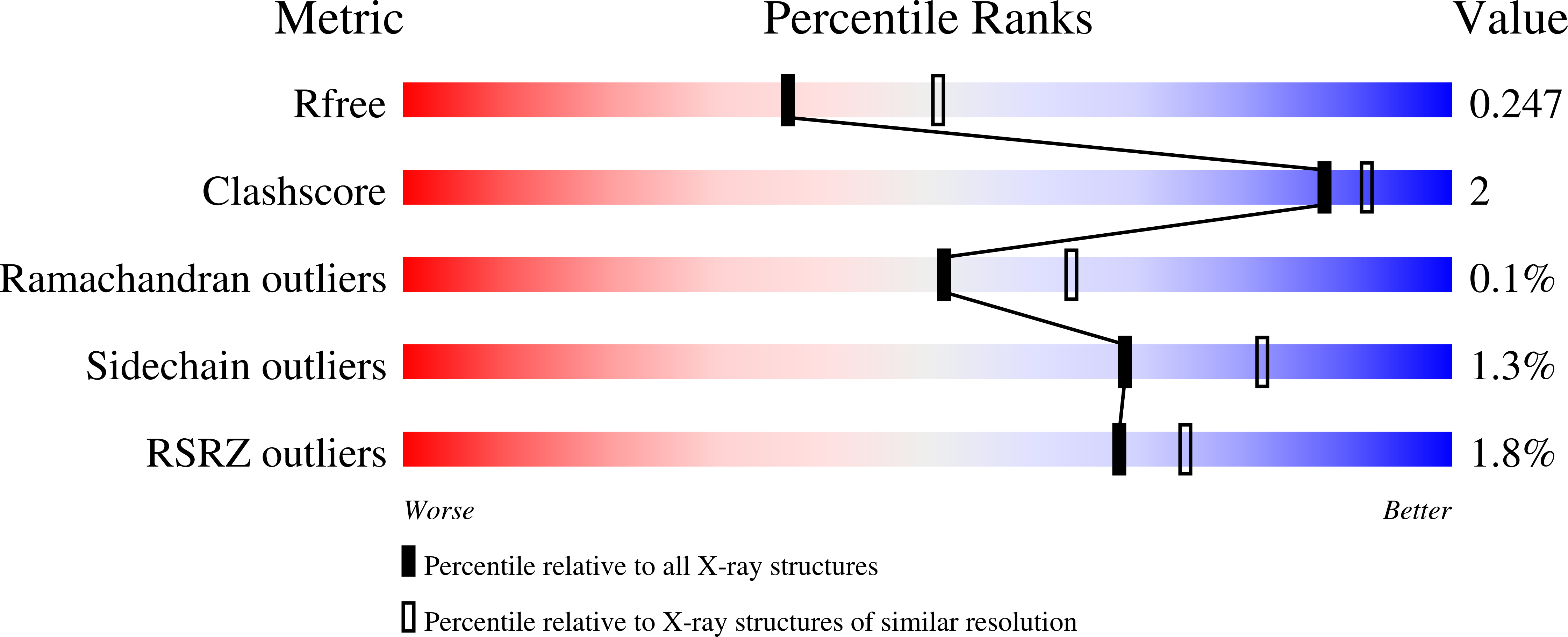
Experimental Data Snapshot
wwPDB Validation 3D Report Full Report
Entity ID: 1 | |||||
|---|---|---|---|---|---|
| Molecule | Chains | Sequence Length | Organism | Details | Image |
| GCN5-related N-acetyltransferase | 173 | Brucella abortus 2308 | Mutation(s): 0 Gene Names: BAB1_1904 EC: 2.3.1 | 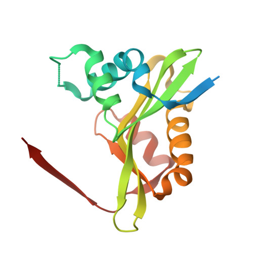 | |
UniProt | |||||
Find proteins for Q2YLP8 (Brucella abortus (strain 2308)) Explore Q2YLP8 Go to UniProtKB: Q2YLP8 | |||||
Entity Groups | |||||
| Sequence Clusters | 30% Identity50% Identity70% Identity90% Identity95% Identity100% Identity | ||||
| UniProt Group | Q2YLP8 | ||||
Sequence AnnotationsExpand | |||||
| |||||
| Length ( Å ) | Angle ( ˚ ) |
|---|---|
| a = 67.02 | α = 87.121 |
| b = 114.9 | β = 76.271 |
| c = 122 | γ = 73.142 |
| Software Name | Purpose |
|---|---|
| XDS | data reduction |
| XSCALE | data scaling |
| PHENIX | refinement |
| PDB_EXTRACT | data extraction |
| MR-Rosetta | phasing |
| PHASER | phasing |
| Coot | model building |
| BUCCANEER | model building |
RCSB PDB (citation) is hosted by
RCSB PDB is a member of the

