8CQB | pdb_00008cqb

Cryo-EM structure of the human GBP1 dimer bound to GDP-AlF3

  • Classification: IMMUNE SYSTEM
  • Organism(s): Homo sapiens
  • Expression System: Escherichia coli
  • Mutation(s): No 

  • Deposited: 2023-03-04 Released: 2024-03-13 
  • Deposition Author(s): Kuhm, T.I., Jakobi, A.J.
  • Funding Organization(s): European Research Council (ERC), Netherlands Organisation for Scientific Research (NWO)

Experimental Data Snapshot

  • Method: ELECTRON MICROSCOPY
  • Resolution: 3.70 Å
  • Aggregation State: PARTICLE 
  • Reconstruction Method: SINGLE PARTICLE 

Starting Models: experimental
View more details

wwPDB Validation   3D Report Full Report


This is version 1.1 of the entry. See complete history


Literature

Structural basis of antimicrobial membrane coat assembly by human GBP1.

Kuhm, T.Taisne, C.de Agrela Pinto, C.Gross, L.Giannopoulou, E.A.Huber, S.T.Pardon, E.Steyaert, J.Tans, S.J.Jakobi, A.J.

(2025) Nat Struct Mol Biol 32: 172-184

  • DOI: https://doi.org/10.1038/s41594-024-01400-9
  • Primary Citation of Related Structures:  
    8CQB

  • PubMed Abstract: 

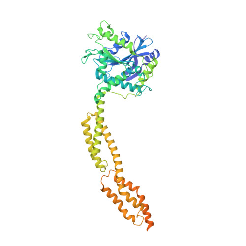
    Guanylate-binding proteins (GBPs) are interferon-inducible guanosine triphosphate hydrolases (GTPases) mediating host defense against intracellular pathogens. Their antimicrobial activity hinges on their ability to self-associate and coat pathogen-associated compartments or cytosolic bacteria. Coat formation depends on GTPase activity but how nucleotide binding and hydrolysis prime coat formation remains unclear. Here, we report the cryo-electron microscopy structure of the full-length human GBP1 dimer in its guanine nucleotide-bound state and describe the molecular ultrastructure of the GBP1 coat on liposomes and bacterial lipopolysaccharide membranes. Conformational changes of the middle and GTPase effector domains expose the isoprenylated C terminus for membrane association. The α-helical middle domains form a parallel, crossover arrangement essential for coat formation and position the extended effector domain for intercalation into the lipopolysaccharide layer of gram-negative membranes. Nucleotide binding and hydrolysis create oligomeric scaffolds with contractile abilities that promote membrane extrusion and fragmentation. Our data offer a structural and mechanistic framework for understanding GBP1 effector functions in intracellular immunity.


  • Organizational Affiliation
    • Department of Bionanoscience, Kavli Insitute of Nanoscience, Delft University of Technology, Delft, The Netherlands.

Macromolecules
Find similar proteins by:  (by identity cutoff)  |  3D Structure
Entity ID: 1
MoleculeChains Sequence LengthOrganismDetailsImage
Guanylate-binding protein 1
A, B
592Homo sapiensMutation(s): 0 
Gene Names: GBP1
EC: 3.6.5 (PDB Primary Data), 3.6.1 (UniProt)
UniProt & NIH Common Fund Data Resources
Find proteins for P32455 (Homo sapiens)
Explore P32455 
Go to UniProtKB:  P32455
PHAROS:  P32455
GTEx:  ENSG00000117228 
Entity Groups  
Sequence Clusters30% Identity50% Identity70% Identity90% Identity95% Identity100% Identity
UniProt GroupP32455
Sequence Annotations
Expand
  • Reference Sequence
Small Molecules
Ligands 3 Unique
IDChains Name / Formula / InChI Key2D Diagram3D Interactions
GDP (Subject of Investigation/LOI)
Query on GDP

Download Ideal Coordinates CCD File 
C [auth A],
F [auth B]
GUANOSINE-5'-DIPHOSPHATE
C10 H15 N5 O11 P2
QGWNDRXFNXRZMB-UUOKFMHZSA-N
AF3 (Subject of Investigation/LOI)
Query on AF3

Download Ideal Coordinates CCD File 
D [auth A],
G [auth B]
ALUMINUM FLUORIDE
Al F3
KLZUFWVZNOTSEM-UHFFFAOYSA-K
MG (Subject of Investigation/LOI)
Query on MG

Download Ideal Coordinates CCD File 
E [auth A],
H [auth B]
MAGNESIUM ION
Mg
JLVVSXFLKOJNIY-UHFFFAOYSA-N
Experimental Data & Validation

Experimental Data

  • Method: ELECTRON MICROSCOPY
  • Resolution: 3.70 Å
  • Aggregation State: PARTICLE 
  • Reconstruction Method: SINGLE PARTICLE 
EM Software:
TaskSoftware PackageVersion
RECONSTRUCTIONcryoSPARC3.3
MODEL REFINEMENTREFMAC5

Structure Validation

View Full Validation Report


Entry History & Funding Information

Deposition Data


Funding OrganizationLocationGrant Number
European Research Council (ERC)European Union852880
Netherlands Organisation for Scientific Research (NWO)NetherlandsNWO.STU.018-2.007

Revision History  (Full details and data files)

  • Version 1.0: 2024-03-13
    Type: Initial release
  • Version 1.1: 2025-01-29
    Changes: Advisory, Data collection, Database references, Derived calculations, Structure summary