8WTB

Crystal structure of McsA/McsB complex truncated by chymotrypsin


Experimental Data Snapshot

  • Method: X-RAY DIFFRACTION
  • Resolution: 2.90 Å
  • R-Value Free: 0.259 
  • R-Value Work: 0.205 
  • R-Value Observed: 0.208 

wwPDB Validation   3D Report Full Report


This is version 1.0 of the entry. See complete history


Literature

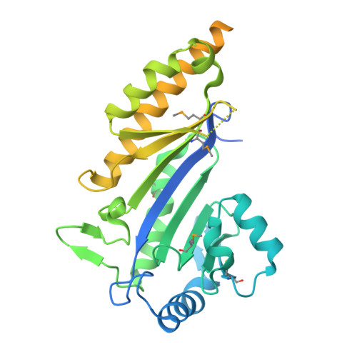
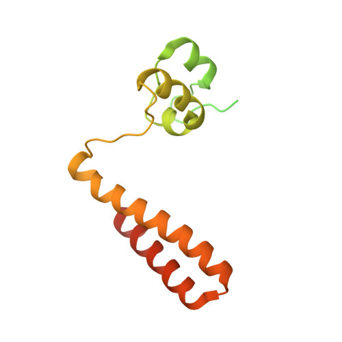
Structural insights into the regulation of protein-arginine kinase McsB by McsA.

Arifuzzaman, M.Kwon, E.Kim, D.Y.

(2024) Proc Natl Acad Sci U S A 121: e2320312121-e2320312121

  • DOI: https://doi.org/10.1073/pnas.2320312121
  • Primary Citation of Related Structures:  
    8WTB, 8WTC

  • PubMed Abstract: 

    In gram-positive bacteria, phosphorylated arginine functions as a protein degradation signal in a similar manner as ubiquitin in eukaryotes. The protein-arginine phosphorylation is mediated by the McsAB complex, where McsB possesses kinase activity and McsA modulates McsB activity. Although mcsA and mcsB are regulated within the same operon, the role of McsA in kinase activity has not yet been clarified. In this study, we determined the molecular mechanism by which McsA regulates kinase activity. The crystal structure of the McsAB complex shows that McsA binds to the McsB kinase domain through a second zinc-coordination domain and the subsequent loop region. This binding activates McsB kinase activity by rearranging the catalytic site, preventing McsB self-assembly, and enhancing stoichiometric substrate binding. The first zinc-coordination and coiled-coil domains of McsA further activate McsB by reassembling the McsAB oligomer. These results demonstrate that McsA is the regulatory subunit for the reconstitution of the protein-arginine kinase holoenzyme. This study provides structural insight into how protein-arginine kinase directs the cellular protein degradation system.


  • Organizational Affiliation

    College of Pharmacy, Yeungnam University, Gyeongsan 38541, Republic of Korea.


Macromolecules
Find similar proteins by:  (by identity cutoff)  |  3D Structure
Entity ID: 1
MoleculeChains Sequence LengthOrganismDetailsImage
Protein-arginine kinase
A, C
365Bacillus subtilis subsp. subtilis str. 168Mutation(s): 0 
Gene Names: mcsB
UniProt
Find proteins for P37570 (Bacillus subtilis (strain 168))
Explore P37570 
Go to UniProtKB:  P37570
Entity Groups  
Sequence Clusters30% Identity50% Identity70% Identity90% Identity95% Identity100% Identity
UniProt GroupP37570
Sequence Annotations
Expand
  • Reference Sequence
Find similar proteins by:  (by identity cutoff)  |  3D Structure
Entity ID: 2
MoleculeChains Sequence LengthOrganismDetailsImage
Protein-arginine kinase activator protein
B, D
187Bacillus subtilis subsp. subtilis str. 168Mutation(s): 0 
Gene Names: mcsA
UniProt
Find proteins for P37569 (Bacillus subtilis (strain 168))
Explore P37569 
Go to UniProtKB:  P37569
Entity Groups  
Sequence Clusters30% Identity50% Identity70% Identity90% Identity95% Identity100% Identity
UniProt GroupP37569
Sequence Annotations
Expand
  • Reference Sequence
Small Molecules
Modified Residues  1 Unique
IDChains TypeFormula2D DiagramParent
MSE
Query on MSE
A, C
L-PEPTIDE LINKINGC5 H11 N O2 SeMET
Experimental Data & Validation

Experimental Data

  • Method: X-RAY DIFFRACTION
  • Resolution: 2.90 Å
  • R-Value Free: 0.259 
  • R-Value Work: 0.205 
  • R-Value Observed: 0.208 
  • Space Group: C 1 2 1
Unit Cell:
Length ( Å )Angle ( ˚ )
a = 179.869α = 90
b = 75.59β = 99.78
c = 79.19γ = 90
Software Package:
Software NamePurpose
PHENIXrefinement
MOSFLMdata reduction
Aimlessdata scaling
PHASERphasing

Structure Validation

View Full Validation Report


Entry History & Funding Information

Deposition Data


Funding OrganizationLocationGrant Number
Not funded--

Revision History  (Full details and data files)

  • Version 1.0: 2024-05-08
    Type: Initial release