9IY2 | pdb_00009iy2

Immune complex of HEV-E2s, nAb 8C11 and nAb 8H3


Experimental Data Snapshot

  • Method: X-RAY DIFFRACTION
  • Resolution: 3.48 Å
  • R-Value Free: 
    0.269 (Depositor), 0.268 (DCC) 
  • R-Value Work: 
    0.223 (Depositor), 0.229 (DCC) 
  • R-Value Observed: 
    0.226 (Depositor) 

Starting Models: experimental
View more details

wwPDB Validation   3D Report Full Report


This is version 1.3 of the entry. See complete history


Literature

Structural basis for the synergetic neutralization of hepatitis E virus by antibody-antibody interaction.

Zheng, M.Zhou, L.Huang, Y.Zhang, X.Yu, Z.Yang, C.Chen, Y.Ying, D.Wang, H.Chen, Z.Liu, C.Tang, Z.Wang, S.Wang, K.Yang, K.Lin, Y.Li, T.Zheng, Q.Zheng, Z.Zhang, J.Yu, H.Li, S.Gu, Y.Xia, N.

(2024) Proc Natl Acad Sci U S A 121: e2408585121-e2408585121

  • DOI: https://doi.org/10.1073/pnas.2408585121
  • Primary Citation of Related Structures:  
    9IY0, 9IY2

  • PubMed Abstract: 

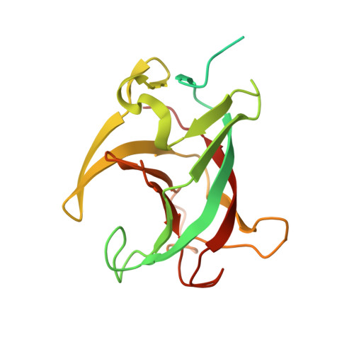
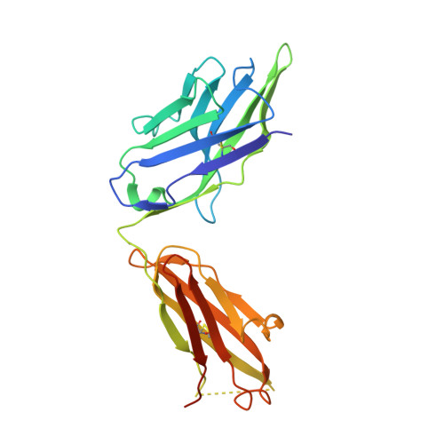
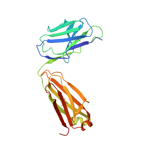
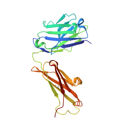
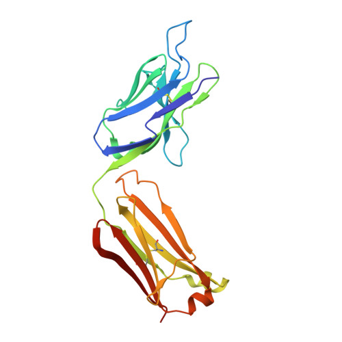
    Neutralizing antibodies (nAbs) play a crucial role in virology, antibody drug development, and vaccine research. In this study, we investigated the synergistic effect of two hepatitis E virus (HEV) nAbs, 8H3, and 8C11, which have exhibited enhanced neutralizing activity in a rhesus monkey model. We presented crystal structures of 8H3 Fab alone and a triple complex of 8C11 Fab and 8H3 Fab simultaneously binding to the HEV E2s protein (8C11:E2s:8H3). Through structural analysis, we identified critical binding sites and fully elucidated the binding footprints of nAb 8H3 in the 8C11:E2s:8H3 complex using site-directed mutagenesis, pinpointing Ile 529, Glu 549, Lys 554, and Ser 566 in the E2s domain, and K66H, S67H, D88H in the 8C11 heavy chain. Interestingly, the synergetic enhancement of 8C11 to 8H3 converted to an antagonistic effect when 8C11 bound to E2s with pretreatment of 8H3, indicating a unidirectional synergistic effect associated with the sequence of antibody involvement. We demonstrated this phenomenon through structural comparisons of E2s:8C11 vs. 8C11:E2s:8H3 crystal structures and molecular dynamics simulations, found that Ile 529 played a key role in the synergistic interplay between these two nAbs. The two-antibody combination showed a more potent antibody-imposed physical disruption mechanism and enhanced coneutralization in an authentic HEV-based cell model. Our study suggests a strategy for synergistic antibody cocktail design with antibody-antibody side-by-side interaction.


  • Organizational Affiliation
    • State Key Laboratory of Vaccines for Infectious Diseases, Xiang An Biomedicine Laboratory, Department of Laboratory Medicine, School of Public Health, School of Life Sciences, Xiamen University, Xiamen 361102, China.

Macromolecules
Find similar proteins by:  (by identity cutoff)  |  3D Structure
Entity ID: 1
MoleculeChains Sequence LengthOrganismDetailsImage
Secreted protein ORF2A,
D [auth B]
214Hepatitis E virus (strain Pakistan)Mutation(s): 0 
Gene Names: ORF2
UniProt
Find proteins for P33426 (Hepatitis E virus genotype 1 (isolate Human/Pakistan/Sar-55))
Explore P33426 
Go to UniProtKB:  P33426
Entity Groups  
Sequence Clusters30% Identity50% Identity70% Identity90% Identity95% Identity100% Identity
UniProt GroupP33426
Sequence Annotations
Expand
  • Reference Sequence
Find similar proteins by:  (by identity cutoff)  |  3D Structure
Entity ID: 2
MoleculeChains Sequence LengthOrganismDetailsImage
Heavy Chain of mAb 8C11B [auth H],
F [auth D]
230Mus sp.Mutation(s): 0 
Entity Groups  
Sequence Clusters30% Identity50% Identity70% Identity90% Identity95% Identity100% Identity
Sequence Annotations
Expand
  • Reference Sequence
Find similar proteins by:  (by identity cutoff)  |  3D Structure
Entity ID: 3
MoleculeChains Sequence LengthOrganismDetailsImage
Light Chain of mAb 8C11C [auth L],
E [auth C]
214Mus sp.Mutation(s): 0 
Entity Groups  
Sequence Clusters30% Identity50% Identity70% Identity90% Identity95% Identity100% Identity
Sequence Annotations
Expand
  • Reference Sequence
Find similar proteins by:  (by identity cutoff)  |  3D Structure
Entity ID: 4
MoleculeChains Sequence LengthOrganismDetailsImage
Heavy Chain of mAb 8H3G [auth E],
I [auth G]
221Mus sp.Mutation(s): 0 
Entity Groups  
Sequence Clusters30% Identity50% Identity70% Identity90% Identity95% Identity100% Identity
Sequence Annotations
Expand
  • Reference Sequence
Find similar proteins by:  (by identity cutoff)  |  3D Structure
Entity ID: 5
MoleculeChains Sequence LengthOrganismDetailsImage
Light Chain of mAb 8H3H [auth F],
J [auth I]
219Mus sp.Mutation(s): 0 
Entity Groups  
Sequence Clusters30% Identity50% Identity70% Identity90% Identity95% Identity100% Identity
Sequence Annotations
Expand
  • Reference Sequence
Experimental Data & Validation

Experimental Data

  • Method: X-RAY DIFFRACTION
  • Resolution: 3.48 Å
  • R-Value Free:  0.269 (Depositor), 0.268 (DCC) 
  • R-Value Work:  0.223 (Depositor), 0.229 (DCC) 
  • R-Value Observed: 0.226 (Depositor) 
Space Group: P 1 21 1
Unit Cell:
Length ( Å )Angle ( ˚ )
a = 71.465α = 90
b = 248.941β = 94.07
c = 76.841γ = 90
Software Package:
Software NamePurpose
PHENIXrefinement
HKL-2000data reduction
HKL-2000data scaling
PHASERphasing

Structure Validation

View Full Validation Report


Entry History & Funding Information

Deposition Data


Funding OrganizationLocationGrant Number
National Science Foundation (NSF, China)China--

Revision History  (Full details and data files)

  • Version 1.0: 2024-08-28
    Type: Initial release
  • Version 1.1: 2024-09-04
    Changes: Database references
  • Version 1.2: 2024-10-30
    Changes: Structure summary
  • Version 1.3: 2025-09-10
    Changes: Database references