1RKY

PPLO + Xe


Experimental Data Snapshot

  • Method: X-RAY DIFFRACTION
  • Resolution: 1.68 Å
  • R-Value Free: 0.182 
  • R-Value Work: 0.151 
  • R-Value Observed: 0.151 

wwPDB Validation   3D Report Full Report


Ligand Structure Quality Assessment 


This is version 2.1 of the entry. See complete history


Literature

Using Xenon as a Probe for Dioxygen-binding Sites in Copper Amine Oxidases

Duff, A.P.Trambaiolo, D.M.Cohen, A.E.Ellis, P.J.Juda, G.A.Shepard, E.M.Langley, D.B.Dooley, D.M.Freeman, H.C.Guss, J.M.

(2004) J Mol Biol 344: 599-607

  • DOI: https://doi.org/10.1016/j.jmb.2004.09.075
  • Primary Citation of Related Structures:  
    1RJO, 1RKY, 1W2Z

  • PubMed Abstract: 

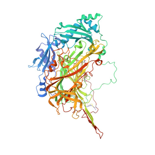
    Potential dioxygen-binding sites in three Cu amine oxidases have been investigated by recording X-ray diffraction data at 1.7-2.2A resolution for crystals under a high pressure of xenon gas. Electron-density difference maps and crystallographic refinement provide unequivocal evidence for a number of Xe-binding sites in each enzyme. Only one of these sites is present in all three Cu amine oxidases studied. Structural changes elsewhere in the protein molecules are insignificant. The results illustrate the use of xenon as a probe for cavities, in which a protein may accommodate a dioxygen molecule. The finding of a potential dioxygen-binding cavity close to the active site of Cu amine oxidases may be relevant to the function of the enzymes, since the formation of a transient protein-dioxygen complex is a likely step in the catalytic mechanism. No evidence was found for xenon binding in a region of the molecule that was previously identified in two other Cu amine oxidases as a potential transient dioxygen-binding site.


  • Organizational Affiliation

    School of Molecular and Microbial Biosciences, University of Sydney, NSW 2006, Australia.


Macromolecules
Find similar proteins by:  (by identity cutoff)  |  3D Structure
Entity ID: 1
MoleculeChains Sequence LengthOrganismDetailsImage
lysyl oxidase747Komagataella pastorisMutation(s): 1 
Gene Names: ATCC 28485
EC: 1.4.3.13
UniProt
Find proteins for Q96X16 (Komagataella pastoris)
Explore Q96X16 
Go to UniProtKB:  Q96X16
Entity Groups  
Sequence Clusters30% Identity50% Identity70% Identity90% Identity95% Identity100% Identity
UniProt GroupQ96X16
Sequence Annotations
Expand
  • Reference Sequence
Oligosaccharides

Help

Entity ID: 2
MoleculeChains Length2D Diagram Glycosylation3D Interactions
beta-D-mannopyranose-(1-4)-2-acetamido-2-deoxy-beta-D-glucopyranose-(1-4)-2-acetamido-2-deoxy-beta-D-glucopyranose
B
3N-Glycosylation
Glycosylation Resources
GlyTouCan:  G15407YE
GlyCosmos:  G15407YE
GlyGen:  G15407YE
Small Molecules
Ligands 7 Unique
IDChains Name / Formula / InChI Key2D Diagram3D Interactions
NAG
Query on NAG

Download Ideal Coordinates CCD File 
C [auth A],
D [auth A],
E [auth A],
F [auth A]
2-acetamido-2-deoxy-beta-D-glucopyranose
C8 H15 N O6
OVRNDRQMDRJTHS-FMDGEEDCSA-N
XE
Query on XE

Download Ideal Coordinates CCD File 
AA [auth A]
T [auth A]
U [auth A]
V [auth A]
W [auth A]
AA [auth A],
T [auth A],
U [auth A],
V [auth A],
W [auth A],
X [auth A],
Y [auth A],
Z [auth A]
XENON
Xe
FHNFHKCVQCLJFQ-UHFFFAOYSA-N
IMD
Query on IMD

Download Ideal Coordinates CCD File 
S [auth A]IMIDAZOLE
C3 H5 N2
RAXXELZNTBOGNW-UHFFFAOYSA-O
CU
Query on CU

Download Ideal Coordinates CCD File 
G [auth A]COPPER (II) ION
Cu
JPVYNHNXODAKFH-UHFFFAOYSA-N
CA
Query on CA

Download Ideal Coordinates CCD File 
H [auth A],
I [auth A]
CALCIUM ION
Ca
BHPQYMZQTOCNFJ-UHFFFAOYSA-N
CL
Query on CL

Download Ideal Coordinates CCD File 
M [auth A]
N [auth A]
O [auth A]
P [auth A]
Q [auth A]
M [auth A],
N [auth A],
O [auth A],
P [auth A],
Q [auth A],
R [auth A]
CHLORIDE ION
Cl
VEXZGXHMUGYJMC-UHFFFAOYSA-M
MG
Query on MG

Download Ideal Coordinates CCD File 
J [auth A],
K [auth A],
L [auth A]
MAGNESIUM ION
Mg
JLVVSXFLKOJNIY-UHFFFAOYSA-N
Modified Residues  1 Unique
IDChains TypeFormula2D DiagramParent
TPQ
Query on TPQ
A
L-PEPTIDE LINKINGC9 H9 N O5TYR
Experimental Data & Validation

Experimental Data

  • Method: X-RAY DIFFRACTION
  • Resolution: 1.68 Å
  • R-Value Free: 0.182 
  • R-Value Work: 0.151 
  • R-Value Observed: 0.151 
  • Space Group: C 1 2 1
Unit Cell:
Length ( Å )Angle ( ˚ )
a = 140.144α = 90
b = 66.702β = 119.24
c = 108.62γ = 90
Software Package:
Software NamePurpose
REFMACrefinement
MOSFLMdata reduction
CCP4data scaling
MOLREPphasing

Structure Validation

View Full Validation Report


Ligand Structure Quality Assessment 


Entry History 

Deposition Data

Revision History  (Full details and data files)

  • Version 1.0: 2004-12-07
    Type: Initial release
  • Version 1.1: 2008-04-29
    Changes: Version format compliance
  • Version 1.2: 2011-07-13
    Changes: Derived calculations, Version format compliance
  • Version 2.0: 2020-07-29
    Type: Remediation
    Reason: Carbohydrate remediation
    Changes: Advisory, Atomic model, Data collection, Database references, Derived calculations, Structure summary
  • Version 2.1: 2023-10-25
    Changes: Advisory, Data collection, Database references, Refinement description, Structure summary