4CBJ

The c-ring ion binding site of the ATP synthase from Bacillus pseudofirmus OF4 is adapted to alkaliphilic cell physiology


Experimental Data Snapshot

  • Method: X-RAY DIFFRACTION
  • Resolution: 2.80 Å
  • R-Value Free: 0.288 
  • R-Value Work: 0.235 
  • R-Value Observed: 0.238 

wwPDB Validation   3D Report Full Report


Ligand Structure Quality Assessment 


This is version 1.2 of the entry. See complete history


Literature

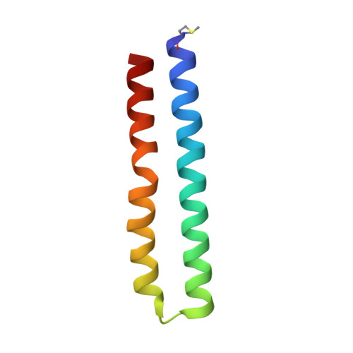
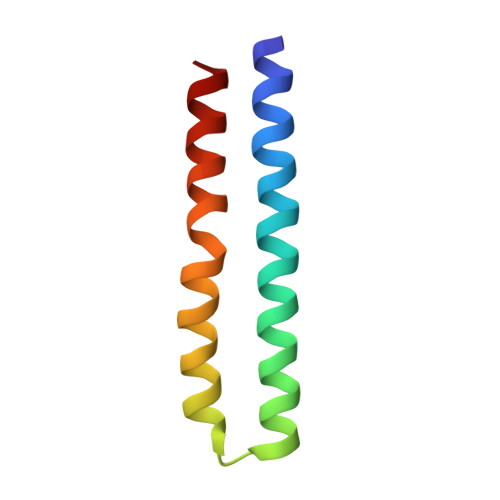
The C-Ring Ion-Binding Site of the ATP Synthase from Bacillus Pseudofirmus of4 is Adapted to Alkaliphilic Lifestyle.

Preiss, L.Langer, J.D.Hicks, D.B.Liu, J.Yildiz, O.Krulwich, T.A.Meier, T.

(2014) Mol Microbiol 92: 973

  • DOI: https://doi.org/10.1111/mmi.12605
  • Primary Citation of Related Structures:  
    4CBJ, 4CBK

  • PubMed Abstract: 

    In the c-ring rotor of ATP synthases ions are shuttled across the membrane during ATP synthesis by a unique rotary mechanism. We investigated characteristics of the c-ring from the alkaliphile Bacillus pseudofirmus OF4 with respect to evolutionary adaptations to operate with protons at high environmental pH. The X-ray structures of the wild-type c13 ring at pH 9.0 and a 'neutralophile-like' mutant (P51A) at pH 4.4, at 2.4 and 2.8 Å resolution, respectively, reveal a dependency of the conformation and protonation state of the proton-binding glutamate (E(54) ) on environmental hydrophobicity. Faster labelling kinetics with the inhibitor dicyclohexylcarbodiimide (DCCD) demonstrate a greater flexibility of E(54) in the mutant due to reduced water occupancy within the H(+) binding site. A second 'neutralophile-like' mutant (V21N) shows reduced growth at high pH, which is explained by restricted conformational freedom of the mutant's E(54) carboxylate. The study directly connects subtle structural adaptations of the c-ring ion binding site to in vivo effects of alkaliphile cell physiology.


  • Organizational Affiliation

    Department of Structural Biology, Max Planck Institute of Biophysics, Max-von-Laue-Str. 3, 60438, Frankfurt am Main, Germany.


Macromolecules
Find similar proteins by:  (by identity cutoff)  |  3D Structure
Entity ID: 1
MoleculeChains Sequence LengthOrganismDetailsImage
ATP SYNTHASE SUBUNIT C
A, B, I, J, K
A, B, I, J, K, L, M
69Alkalihalophilus pseudofirmus OF4Mutation(s): 0 
Membrane Entity: Yes 
UniProt
Find proteins for P22483 (Alkalihalophilus pseudofirmus (strain ATCC BAA-2126 / JCM 17055 / OF4))
Explore P22483 
Go to UniProtKB:  P22483
Entity Groups  
Sequence Clusters30% Identity50% Identity70% Identity90% Identity95% Identity100% Identity
UniProt GroupP22483
Sequence Annotations
Expand
  • Reference Sequence
Find similar proteins by:  (by identity cutoff)  |  3D Structure
Entity ID: 2
MoleculeChains Sequence LengthOrganismDetailsImage
ATP SYNTHASE SUBUNIT C
C, D, E, F, G
C, D, E, F, G, H
69Alkalihalophilus pseudofirmus OF4Mutation(s): 0 
Membrane Entity: Yes 
UniProt
Find proteins for P22483 (Alkalihalophilus pseudofirmus (strain ATCC BAA-2126 / JCM 17055 / OF4))
Explore P22483 
Go to UniProtKB:  P22483
Entity Groups  
Sequence Clusters30% Identity50% Identity70% Identity90% Identity95% Identity100% Identity
UniProt GroupP22483
Sequence Annotations
Expand
  • Reference Sequence
Small Molecules
Ligands 3 Unique
IDChains Name / Formula / InChI Key2D Diagram3D Interactions
LMT
Query on LMT

Download Ideal Coordinates CCD File 
Z [auth J]DODECYL-BETA-D-MALTOSIDE
C24 H46 O11
NLEBIOOXCVAHBD-QKMCSOCLSA-N
DPV
Query on DPV

Download Ideal Coordinates CCD File 
AA [auth K]
CA [auth L]
DA [auth M]
N [auth A]
P [auth B]
AA [auth K],
CA [auth L],
DA [auth M],
N [auth A],
P [auth B],
Q [auth C],
R [auth D],
T [auth E],
U [auth F],
V [auth G],
W [auth H],
X [auth I],
Y [auth J]
dodecyl 2-(trimethylammonio)ethyl phosphate
C17 H38 N O4 P
QBHFVMDLPTZDOI-UHFFFAOYSA-N
TAM
Query on TAM

Download Ideal Coordinates CCD File 
BA [auth K],
O [auth A],
S [auth D]
TRIS(HYDROXYETHYL)AMINOMETHANE
C7 H17 N O3
GKODZWOPPOTFGA-UHFFFAOYSA-N
Modified Residues  1 Unique
IDChains TypeFormula2D DiagramParent
FME
Query on FME
A, B, I, J, K
A, B, I, J, K, L, M
L-PEPTIDE LINKINGC6 H11 N O3 SMET
Experimental Data & Validation

Experimental Data

  • Method: X-RAY DIFFRACTION
  • Resolution: 2.80 Å
  • R-Value Free: 0.288 
  • R-Value Work: 0.235 
  • R-Value Observed: 0.238 
  • Space Group: P 1 21 1
Unit Cell:
Length ( Å )Angle ( ˚ )
a = 73.94α = 90
b = 98.33β = 104.28
c = 121.93γ = 90
Software Package:
Software NamePurpose
PHENIXrefinement
XDSdata reduction
XSCALEdata scaling
PHASERphasing

Structure Validation

View Full Validation Report


Ligand Structure Quality Assessment 


Entry History 

Deposition Data

Revision History  (Full details and data files)

  • Version 1.0: 2014-05-28
    Type: Initial release
  • Version 1.1: 2014-06-11
    Changes: Database references
  • Version 1.2: 2023-12-20
    Changes: Data collection, Database references, Derived calculations, Other, Refinement description