4KYI

Crystal structure of the phospholipase VipD from Legionella pneumophila in complex with the human GTPase Rab5


Experimental Data Snapshot

  • Method: X-RAY DIFFRACTION
  • Resolution: 3.08 Å
  • R-Value Free: 0.280 
  • R-Value Work: 0.233 
  • R-Value Observed: 0.235 

wwPDB Validation   3D Report Full Report


Ligand Structure Quality Assessment 


This is version 1.2 of the entry. See complete history


Literature

Structural basis for the recruitment and activation of the Legionella phospholipase VipD by the host GTPase Rab5.

Lucas, M.Gaspar, A.H.Pallara, C.Rojas, A.L.Fernandez-Recio, J.Machner, M.P.Hierro, A.

(2014) Proc Natl Acad Sci U S A 111: E3514-E3523

  • DOI: https://doi.org/10.1073/pnas.1405391111
  • Primary Citation of Related Structures:  
    4KYI

  • PubMed Abstract: 

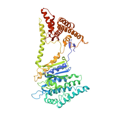
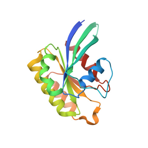
    A challenge for microbial pathogens is to assure that their translocated effector proteins target only the correct host cell compartment during infection. The Legionella pneumophila effector vacuolar protein sorting inhibitor protein D (VipD) localizes to early endosomal membranes and alters their lipid and protein composition, thereby protecting the pathogen from endosomal fusion. This process requires the phospholipase A1 (PLA1) activity of VipD that is triggered specifically on VipD binding to the host cell GTPase Rab5, a key regulator of endosomes. Here, we present the crystal structure of VipD in complex with constitutively active Rab5 and reveal the molecular mechanism underlying PLA1 activation. An active site-obstructing loop that originates from the C-terminal domain of VipD is repositioned on Rab5 binding, thereby exposing the catalytic pocket within the N-terminal PLA1 domain. Substitution of amino acid residues located within the VipD-Rab5 interface prevented Rab5 binding and PLA1 activation and caused a failure of VipD mutant proteins to target to Rab5-enriched endosomal structures within cells. Experimental and computational analyses confirmed an extended VipD-binding interface on Rab5, explaining why this L. pneumophila effector can compete with cellular ligands for Rab5 binding. Together, our data explain how the catalytic activity of a microbial effector can be precisely linked to its subcellular localization.


  • Organizational Affiliation

    Structural Biology Unit, Center for Cooperative Research in Biosciences, 48160 Derio, Spain;


Macromolecules
Find similar proteins by:  (by identity cutoff)  |  3D Structure
Entity ID: 1
MoleculeChains Sequence LengthOrganismDetailsImage
VipD
A, C, E, G
551Legionella pneumophila subsp. pneumophila str. Philadelphia 1Mutation(s): 0 
Gene Names: vipDlpg2831
UniProt
Find proteins for Q5ZRP9 (Legionella pneumophila subsp. pneumophila (strain Philadelphia 1 / ATCC 33152 / DSM 7513))
Explore Q5ZRP9 
Go to UniProtKB:  Q5ZRP9
Entity Groups  
Sequence Clusters30% Identity50% Identity70% Identity90% Identity95% Identity100% Identity
UniProt GroupQ5ZRP9
Sequence Annotations
Expand
  • Reference Sequence
Find similar proteins by:  (by identity cutoff)  |  3D Structure
Entity ID: 2
MoleculeChains Sequence LengthOrganismDetailsImage
Ras-related protein Rab-5C
B, D, F, H
170Homo sapiensMutation(s): 1 
Gene Names: RAB5CRABL
Membrane Entity: Yes 
UniProt & NIH Common Fund Data Resources
Find proteins for P51148 (Homo sapiens)
Explore P51148 
Go to UniProtKB:  P51148
PHAROS:  P51148
GTEx:  ENSG00000108774 
Entity Groups  
Sequence Clusters30% Identity50% Identity70% Identity90% Identity95% Identity100% Identity
UniProt GroupP51148
Sequence Annotations
Expand
  • Reference Sequence
Small Molecules
Ligands 4 Unique
IDChains Name / Formula / InChI Key2D Diagram3D Interactions
GNP
Query on GNP

Download Ideal Coordinates CCD File 
M [auth B],
Q [auth D],
U [auth F],
X [auth H]
PHOSPHOAMINOPHOSPHONIC ACID-GUANYLATE ESTER
C10 H17 N6 O13 P3
UQABYHGXWYXDTK-UUOKFMHZSA-N
GOL
Query on GOL

Download Ideal Coordinates CCD File 
K [auth A],
L [auth A],
T [auth E]
GLYCEROL
C3 H8 O3
PEDCQBHIVMGVHV-UHFFFAOYSA-N
EDO
Query on EDO

Download Ideal Coordinates CCD File 
I [auth A]
J [auth A]
N [auth B]
O [auth B]
S [auth E]
I [auth A],
J [auth A],
N [auth B],
O [auth B],
S [auth E],
W [auth G]
1,2-ETHANEDIOL
C2 H6 O2
LYCAIKOWRPUZTN-UHFFFAOYSA-N
MG
Query on MG

Download Ideal Coordinates CCD File 
P [auth B],
R [auth D],
V [auth F],
Y [auth H]
MAGNESIUM ION
Mg
JLVVSXFLKOJNIY-UHFFFAOYSA-N
Experimental Data & Validation

Experimental Data

  • Method: X-RAY DIFFRACTION
  • Resolution: 3.08 Å
  • R-Value Free: 0.280 
  • R-Value Work: 0.233 
  • R-Value Observed: 0.235 
  • Space Group: P 1
Unit Cell:
Length ( Å )Angle ( ˚ )
a = 94.275α = 76.57
b = 97.976β = 80.71
c = 109.849γ = 78.91
Software Package:
Software NamePurpose
XSCALEdata scaling
PHENIXrefinement
PDB_EXTRACTdata extraction
GDAdata collection
XDSdata reduction
XDSdata scaling
PHASERphasing

Structure Validation

View Full Validation Report


Ligand Structure Quality Assessment 


Entry History 

Deposition Data

Revision History  (Full details and data files)

  • Version 1.0: 2014-08-13
    Type: Initial release
  • Version 1.1: 2014-09-24
    Changes: Database references
  • Version 1.2: 2015-07-29
    Changes: Structure summary