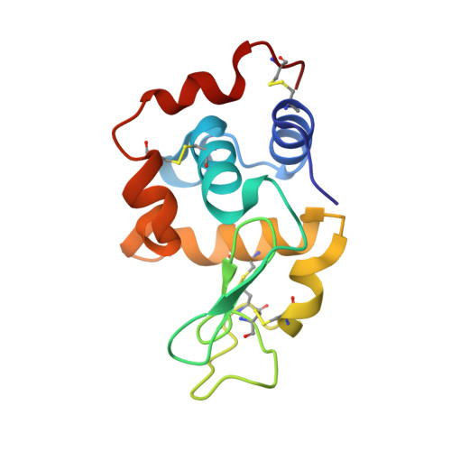
Crystal structure of duck egg lysozyme isoform II (DEL-II).
Langley, D.B., Christ, D.(2018) BMC Struct Biol 18: 10-10
- PubMed: 30134879
- DOI: https://doi.org/10.1186/s12900-018-0090-7
- Primary Citation of Related Structures:
6D9I - PubMed Abstract:
Lysozyme purified from duck eggs (DEL) has long been used as a model antigen as a counterpoint to the enzyme purified from hen eggs (HEL). However, unlike the single C-type variant found in hen eggs, duck eggs contain multiple isoforms: I, II and III. We recently reported the structures of isoforms I and III from Pekin duck (Anas platyrhynchos) and unequivocally determined the sequences of all three isoforms by mass spectrometry. Here we present the crystal structure of isoform II (DEL-II). Lysozyme isoform II was purified from isoforms I and III using ion-exchange and gel-filtration chromatography, then crystallized. X-ray diffraction data were collected to 1.15 Å resolution and the structure of DEL-II was solved by molecular replacement using the structure of DEL-I as the search model. It contains two molecules in the crystallographic asymmetric unit: both molecules display a canonical C-type lysozyme fold and electron density consistent with the expected sequence. The most significant difference between the two molecules concerns different conformations of a surface loop containing one of the expected amino acid differences between the isoforms. The structure of DEL-II supports the primary sequence as elucidated by a combination of amino acid sequencing, DNA sequencing and mass spectrometry, with strong electron density confirming it to be an S37G G71R variant of DEL I, and differing from hen egg lysozyme at a total of 21 amino acid positions.
Organizational Affiliation:
Immunology Division, Garvan Institute of Medical Research, 384 Victoria Road, Darlinghurst, Sydney, NSW, 2010, Australia. d.langley@garvan.org.au.