6J0F

Cryo-EM Structure of an Extracellular Contractile Injection System, PVC sheath/tube terminator in extended state


Experimental Data Snapshot

  • Method: ELECTRON MICROSCOPY
  • Resolution: 3.80 Å
  • Aggregation State: HELICAL ARRAY 
  • Reconstruction Method: SINGLE PARTICLE 

wwPDB Validation   3D Report Full Report


This is version 1.1 of the entry. See complete history


Literature

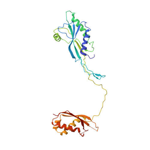
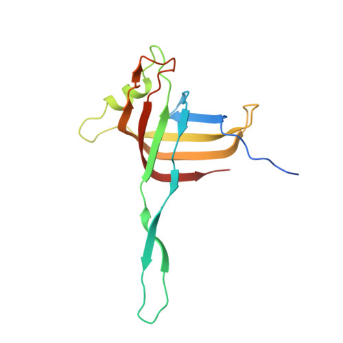
Cryo-EM Structure and Assembly of an Extracellular Contractile Injection System.

Jiang, F.Li, N.Wang, X.Cheng, J.Huang, Y.Yang, Y.Yang, J.Cai, B.Wang, Y.P.Jin, Q.Gao, N.

(2019) Cell 177: 370-383.e15

  • DOI: https://doi.org/10.1016/j.cell.2019.02.020
  • Primary Citation of Related Structures:  
    6J0B, 6J0C, 6J0F, 6J0M, 6J0N

  • PubMed Abstract: 

    Contractile injection systems (CISs) are cell-puncturing nanodevices that share ancestry with contractile tail bacteriophages. Photorhabdus virulence cassette (PVC) represents one group of extracellular CISs that are present in both bacteria and archaea. Here, we report the cryo-EM structure of an intact PVC from P. asymbiotica. This over 10-MDa device resembles a simplified T4 phage tail, containing a hexagonal baseplate complex with six fibers and a capped 117-nanometer sheath-tube trunk. One distinct feature of the PVC is the presence of three variants for both tube and sheath proteins, indicating a functional specialization of them during evolution. The terminal hexameric cap docks onto the topmost layer of the inner tube and locks the outer sheath in pre-contraction state with six stretching arms. Our results on the PVC provide a framework for understanding the general mechanism of widespread CISs and pave the way for using them as delivery tools in biological or therapeutic applications.


  • Organizational Affiliation

    NHC Key Laboratory of Systems Biology of Pathogens, Institute of Pathogen Biology, Chinese Academy of Medical Sciences & Peking Union Medical College, Beijing, PRC.


Macromolecules
Find similar proteins by:  (by identity cutoff)  |  3D Structure
Entity ID: 1
MoleculeChains Sequence LengthOrganismDetailsImage
Pvc16
A, B, C, D, E
A, B, C, D, E, F
293Photorhabdus asymbiotica subsp. asymbiotica ATCC 43949Mutation(s): 0 
Gene Names: PAU_03338PA-RVA20-21-0156
UniProt
Find proteins for B6VNM9 (Photorhabdus asymbiotica subsp. asymbiotica (strain ATCC 43949 / 3105-77))
Explore B6VNM9 
Go to UniProtKB:  B6VNM9
Entity Groups  
Sequence Clusters30% Identity50% Identity70% Identity90% Identity95% Identity100% Identity
UniProt GroupB6VNM9
Sequence Annotations
Expand
  • Reference Sequence
Find similar proteins by:  (by identity cutoff)  |  3D Structure
Entity ID: 2
MoleculeChains Sequence LengthOrganismDetailsImage
Pvc1149Photorhabdus asymbiotica subsp. asymbiotica ATCC 43949Mutation(s): 0 
Gene Names: PAU_03353PA-RVA20-21-0171
UniProt
Find proteins for B6VNP4 (Photorhabdus asymbiotica subsp. asymbiotica (strain ATCC 43949 / 3105-77))
Explore B6VNP4 
Go to UniProtKB:  B6VNP4
Entity Groups  
Sequence Clusters30% Identity50% Identity70% Identity90% Identity95% Identity100% Identity
UniProt GroupB6VNP4
Sequence Annotations
Expand
  • Reference Sequence
Experimental Data & Validation

Experimental Data

  • Method: ELECTRON MICROSCOPY
  • Resolution: 3.80 Å
  • Aggregation State: HELICAL ARRAY 
  • Reconstruction Method: SINGLE PARTICLE 

Structure Validation

View Full Validation Report


Entry History 

Deposition Data

Revision History  (Full details and data files)

  • Version 1.0: 2019-04-10
    Type: Initial release
  • Version 1.1: 2019-04-17
    Changes: Data collection, Database references