


Crystal structure of Mycobacterium tuberculosis dethiobiotin synthetase in complex with fragment degradation product B9D
Thompson, A.P., Polyak, S.W., Wegener, K.L., Bruning, J.B.To be published.
Experimental Data Snapshot
Entity ID: 1 | |||||
|---|---|---|---|---|---|
| Molecule | Chains | Sequence Length | Organism | Details | Image |
| ATP-dependent dethiobiotin synthetase BioD | 233 | Mycobacterium tuberculosis H37Rv | Mutation(s): 0 Gene Names: bioD, Rv1570, MTCY336.33c EC: 6.3.3.3 | 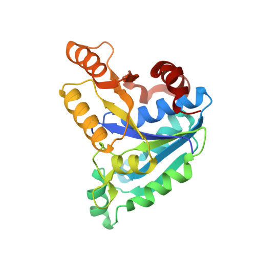 | |
UniProt | |||||
Find proteins for P9WPQ5 (Mycobacterium tuberculosis (strain ATCC 25618 / H37Rv)) Explore P9WPQ5 Go to UniProtKB: P9WPQ5 | |||||
Entity Groups | |||||
| Sequence Clusters | 30% Identity50% Identity70% Identity90% Identity95% Identity100% Identity | ||||
| UniProt Group | P9WPQ5 | ||||
Sequence AnnotationsExpand | |||||
| |||||
| Ligands 3 Unique | |||||
|---|---|---|---|---|---|
| ID | Chains | Name / Formula / InChI Key | 2D Diagram | 3D Interactions | |
| KSJ (Subject of Investigation/LOI) Query on KSJ | E [auth A], I [auth B], M [auth C], Q [auth D] | [(1S,2R)-2-(2-hydroxybenzene-1-carbonyl)cyclopentyl]acetic acid C14 H16 O4 LULBZYDRYZGRST-VHSXEESVSA-N |  | ||
| KSP (Subject of Investigation/LOI) Query on KSP | F [auth A], J [auth B], N [auth C], R [auth D] | [(1R,2S)-2-(2-hydroxybenzene-1-carbonyl)cyclopentyl]acetic acid C14 H16 O4 LULBZYDRYZGRST-ZJUUUORDSA-N |  | ||
| SO4 (Subject of Investigation/LOI) Query on SO4 | G [auth A] H [auth A] K [auth B] L [auth B] O [auth C] | SULFATE ION O4 S QAOWNCQODCNURD-UHFFFAOYSA-L |  | ||
| Length ( Å ) | Angle ( ˚ ) |
|---|---|
| a = 54.61 | α = 90 |
| b = 105.72 | β = 90 |
| c = 154.53 | γ = 90 |
| Software Name | Purpose |
|---|---|
| Aimless | data scaling |
| PHASER | phasing |
| PHENIX | refinement |
| PDB_EXTRACT | data extraction |
| XDS | data reduction |
| Funding Organization | Location | Grant Number |
|---|---|---|
| National Health and Medical Research Council (NHMRC, Australia) | Australia | APP1068885 |


