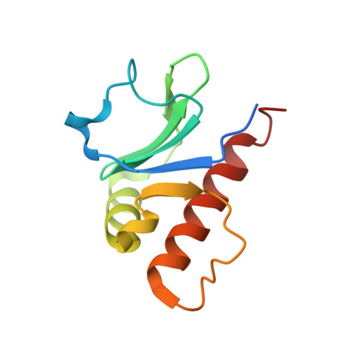
6P5H

Structure of MavC middle insertion domain


Experimental Data Snapshot

  • Method: X-RAY DIFFRACTION
  • Resolution: 1.53 Å
  • R-Value Free: 0.224 
  • R-Value Work: 0.199 
  • R-Value Observed: 0.200 

wwPDB Validation   3D Report Full Report


This is version 1.1 of the entry. See complete history


Literature

Legionella effector MavC targets the Ube2N~Ub conjugate for noncanonical ubiquitination.

Puvar, K.Iyer, S.Fu, J.Kenny, S.Negron Teron, K.I.Luo, Z.Q.Brzovic, P.S.Klevit, R.E.Das, C.

(2020) Nat Commun 11: 2365-2365

  • DOI: https://doi.org/10.1038/s41467-020-16211-x
  • Primary Citation of Related Structures:  
    6P5B, 6P5H, 6ULH, 6UMP, 6UMS

  • PubMed Abstract: 

    The bacterial effector MavC modulates the host immune response by blocking Ube2N activity employing an E1-independent ubiquitin ligation, catalyzing formation of a γ-glutamyl-ε-Lys (Gln40 Ub -Lys92 Ube2N ) isopeptide crosslink using a transglutaminase mechanism. Here we provide biochemical evidence in support of MavC targeting the activated, thioester-linked Ube2N~ubiquitin conjugate, catalyzing an intramolecular transglutamination reaction, covalently crosslinking the Ube2N and Ub subunits effectively inactivating the E2~Ub conjugate. Ubiquitin exhibits weak binding to MavC alone, but shows an increase in affinity when tethered to Ube2N in a disulfide-linked substrate that mimics the charged E2~Ub conjugate. Crystal structures of MavC in complex with the substrate mimic and crosslinked product provide insights into the reaction mechanism and underlying protein dynamics that favor transamidation over deamidation, while revealing a crucial role for the structurally unique insertion domain in substrate recognition. This work provides a structural basis of ubiquitination by transglutamination and identifies this enzyme's true physiological substrate.


  • Organizational Affiliation

    Department of Chemistry, Purdue University, West Lafayette, IN, 47907, USA.


Macromolecules
Find similar proteins by:  (by identity cutoff)  |  3D Structure
Entity ID: 1
MoleculeChains Sequence LengthOrganismDetailsImage
MavC
A, B
105Legionella pneumophilaMutation(s): 0 
Gene Names: C3927_10720
UniProt
Find proteins for Q5ZTL4 (Legionella pneumophila subsp. pneumophila (strain Philadelphia 1 / ATCC 33152 / DSM 7513))
Explore Q5ZTL4 
Go to UniProtKB:  Q5ZTL4
Entity Groups  
Sequence Clusters30% Identity50% Identity70% Identity90% Identity95% Identity100% Identity
UniProt GroupQ5ZTL4
Sequence Annotations
Expand
  • Reference Sequence
Experimental Data & Validation

Experimental Data

  • Method: X-RAY DIFFRACTION
  • Resolution: 1.53 Å
  • R-Value Free: 0.224 
  • R-Value Work: 0.199 
  • R-Value Observed: 0.200 
  • Space Group: P 31
Unit Cell:
Length ( Å )Angle ( ˚ )
a = 52.027α = 90
b = 52.027β = 90
c = 79.753γ = 120
Software Package:
Software NamePurpose
PHENIXrefinement
PDB_EXTRACTdata extraction
HKL-3000data reduction
HKL-3000data scaling
PHASERphasing

Structure Validation

View Full Validation Report


Entry History & Funding Information

Deposition Data


Funding OrganizationLocationGrant Number
National Institutes of Health/National Institute of General Medical Sciences (NIH/NIGMS)United States5R01GM126296-02

Revision History  (Full details and data files)

  • Version 1.0: 2020-05-27
    Type: Initial release
  • Version 1.1: 2023-10-11
    Changes: Data collection, Database references, Refinement description