6TU7

Structure of PfMyoA decorated Plasmodium Act1 filament


Experimental Data Snapshot

  • Method: ELECTRON MICROSCOPY
  • Resolution: 3.10 Å
  • Aggregation State: FILAMENT 
  • Reconstruction Method: HELICAL 

wwPDB Validation   3D Report Full Report


This is version 1.1 of the entry. See complete history


Literature

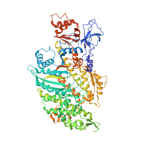
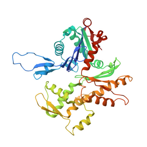
High-resolution structures of malaria parasite actomyosin and actin filaments.

Vahokoski, J.Calder, L.J.Lopez, A.J.Molloy, J.E.Kursula, I.Rosenthal, P.B.

(2022) PLoS Pathog 18: e1010408-e1010408

  • DOI: https://doi.org/10.1371/journal.ppat.1010408
  • Primary Citation of Related Structures:  
    6TU4, 6TU7

  • PubMed Abstract: 

    Malaria is responsible for half a million deaths annually and poses a huge economic burden on the developing world. The mosquito-borne parasites (Plasmodium spp.) that cause the disease depend upon an unconventional actomyosin motor for both gliding motility and host cell invasion. The motor system, often referred to as the glideosome complex, remains to be understood in molecular terms and is an attractive target for new drugs that might block the infection pathway. Here, we present the high-resolution structure of the actomyosin motor complex from Plasmodium falciparum. The complex includes the malaria parasite actin filament (PfAct1) complexed with the class XIV myosin motor (PfMyoA) and its two associated light-chains. The high-resolution core structure reveals the PfAct1:PfMyoA interface in atomic detail, while at lower-resolution, we visualize the PfMyoA light-chain binding region, including the essential light chain (PfELC) and the myosin tail interacting protein (PfMTIP). Finally, we report a bare PfAct1 filament structure at improved resolution.


  • Organizational Affiliation

    Department of Biomedicine, University of Bergen, Bergen, Norway.


Macromolecules
Find similar proteins by:  (by identity cutoff)  |  3D Structure
Entity ID: 1
MoleculeChains Sequence LengthOrganismDetailsImage
Myosin-AA [auth AP1],
F [auth GP1]
820Plasmodium falciparum 3D7Mutation(s): 0 
Gene Names: PF13_0233
UniProt
Find proteins for Q8IDR3 (Plasmodium falciparum (isolate 3D7))
Explore Q8IDR3 
Go to UniProtKB:  Q8IDR3
Entity Groups  
Sequence Clusters30% Identity50% Identity70% Identity90% Identity95% Identity100% Identity
UniProt GroupQ8IDR3
Sequence Annotations
Expand
  • Reference Sequence
Find similar proteins by:  (by identity cutoff)  |  3D Structure
Entity ID: 2
MoleculeChains Sequence LengthOrganismDetailsImage
Actin-1B [auth BP1],
C [auth CP1],
D [auth DP1],
E [auth EP1]
378Plasmodium falciparum 3D7Mutation(s): 0 
Gene Names: PFL2215w
UniProt
Find proteins for Q8I4X0 (Plasmodium falciparum (isolate 3D7))
Explore Q8I4X0 
Go to UniProtKB:  Q8I4X0
Entity Groups  
Sequence Clusters30% Identity50% Identity70% Identity90% Identity95% Identity100% Identity
UniProt GroupQ8I4X0
Sequence Annotations
Expand
  • Reference Sequence
Small Molecules
Ligands 3 Unique
IDChains Name / Formula / InChI Key2D Diagram3D Interactions
9UE
Query on 9UE

Download Ideal Coordinates CCD File 
I [auth BP1],
L [auth CP1],
O [auth DP1],
R [auth EP1]
Jasplakinolide
C36 H45 Br N4 O6
GQWYWHOHRVVHAP-DHKPLNAMSA-N
ADP (Subject of Investigation/LOI)
Query on ADP

Download Ideal Coordinates CCD File 
G [auth BP1],
J [auth CP1],
M [auth DP1],
P [auth EP1]
ADENOSINE-5'-DIPHOSPHATE
C10 H15 N5 O10 P2
XTWYTFMLZFPYCI-KQYNXXCUSA-N
MG (Subject of Investigation/LOI)
Query on MG

Download Ideal Coordinates CCD File 
H [auth BP1],
K [auth CP1],
N [auth DP1],
Q [auth EP1]
MAGNESIUM ION
Mg
JLVVSXFLKOJNIY-UHFFFAOYSA-N
Modified Residues  1 Unique
IDChains TypeFormula2D DiagramParent
SEP
Query on SEP
A [auth AP1],
F [auth GP1]
L-PEPTIDE LINKINGC3 H8 N O6 PSER
Experimental Data & Validation

Experimental Data

  • Method: ELECTRON MICROSCOPY
  • Resolution: 3.10 Å
  • Aggregation State: FILAMENT 
  • Reconstruction Method: HELICAL 
EM Software:
TaskSoftware PackageVersion
RECONSTRUCTIONRELION3.0 beta
MODEL REFINEMENTPHENIX1.16-3549

Structure Validation

View Full Validation Report


Entry History & Funding Information

Deposition Data


Funding OrganizationLocationGrant Number
Norwegian Research CouncilNorway--
The Francis Crick InstituteUnited Kingdom--

Revision History  (Full details and data files)

  • Version 1.0: 2021-01-13
    Type: Initial release
  • Version 1.1: 2022-08-17
    Changes: Database references