6RUP

Human mitochondrial single-stranded DNA binding protein, SSBP1, at 2.1 A resolution - elucidated sequence


Experimental Data Snapshot

  • Method: X-RAY DIFFRACTION
  • Resolution: 2.10 Å
  • R-Value Free: 0.249 
  • R-Value Work: 0.200 
  • R-Value Observed: 0.202 

wwPDB Validation   3D Report Full Report


This is version 1.2 of the entry. See complete history


Literature

Dominant mutations in mtDNA maintenance gene SSBP1 cause optic atrophy and foveopathy.

Piro-Megy, C.Sarzi, E.Tarres-Sole, A.Pequignot, M.Hensen, F.Quiles, M.Manes, G.Chakraborty, A.Senechal, A.Bocquet, B.Cazevieille, C.Roubertie, A.Muller, A.Charif, M.Goudenege, D.Lenaers, G.Wilhelm, H.Kellner, U.Weisschuh, N.Wissinger, B.Zanlonghi, X.Hamel, C.Spelbrink, J.N.Sola, M.Delettre, C.

(2020) J Clin Invest 130: 143-156

  • DOI: https://doi.org/10.1172/JCI128513
  • Primary Citation of Related Structures:  
    6RUP

  • PubMed Abstract: 

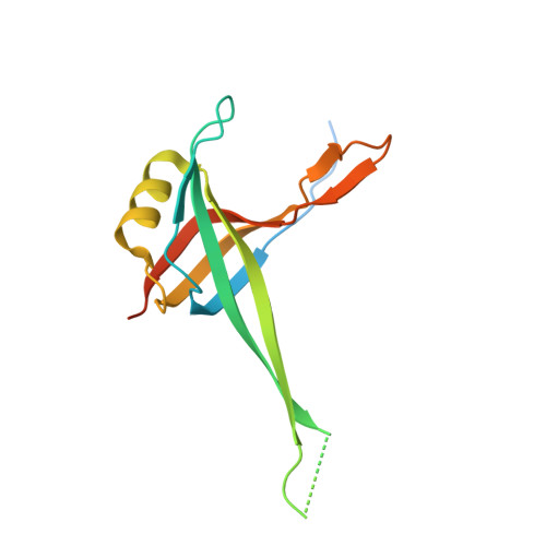
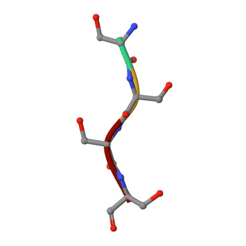
    Mutations in genes encoding components of the mitochondrial DNA (mtDNA) replication machinery cause mtDNA depletion syndromes (MDSs), which associate ocular features with severe neurological syndromes. Here, we identified heterozygous missense mutations in single-strand binding protein 1 (SSBP1) in 5 unrelated families, leading to the R38Q and R107Q amino acid changes in the mitochondrial single-stranded DNA-binding protein, a crucial protein involved in mtDNA replication. All affected individuals presented optic atrophy, associated with foveopathy in half of the cases. To uncover the structural features underlying SSBP1 mutations, we determined a revised SSBP1 crystal structure. Structural analysis suggested that both mutations affect dimer interactions and presumably distort the DNA-binding region. Using patient fibroblasts, we validated that the R38Q variant destabilizes SSBP1 dimer/tetramer formation, affects mtDNA replication, and induces mtDNA depletion. Our study showing that mutations in SSBP1 cause a form of dominant optic atrophy frequently accompanied with foveopathy brings insights into mtDNA maintenance disorders.


  • Organizational Affiliation

    Institute of Neurosciences of Montpellier, INSERM, University of Montpellier, Montpellier, France.


Macromolecules
Find similar proteins by:  (by identity cutoff)  |  3D Structure
Entity ID: 1
MoleculeChains Sequence LengthOrganismDetailsImage
Single-stranded DNA-binding protein, mitochondrial
A, B
142Homo sapiensMutation(s): 0 
Gene Names: SSBP1SSBP
UniProt & NIH Common Fund Data Resources
Find proteins for Q04837 (Homo sapiens)
Explore Q04837 
Go to UniProtKB:  Q04837
PHAROS:  Q04837
GTEx:  ENSG00000106028 
Entity Groups  
Sequence Clusters30% Identity50% Identity70% Identity90% Identity95% Identity100% Identity
UniProt GroupQ04837
Sequence Annotations
Expand
  • Reference Sequence

Find similar proteins by:  Sequence   |   3D Structure  

Entity ID: 2
MoleculeChains Sequence LengthOrganismDetailsImage
SER-SER-SER-SER4Homo sapiensMutation(s): 0 
Sequence Annotations
Expand
  • Reference Sequence
Experimental Data & Validation

Experimental Data

  • Method: X-RAY DIFFRACTION
  • Resolution: 2.10 Å
  • R-Value Free: 0.249 
  • R-Value Work: 0.200 
  • R-Value Observed: 0.202 
  • Space Group: P 41 21 2
Unit Cell:
Length ( Å )Angle ( ˚ )
a = 51.04α = 90
b = 51.04β = 90
c = 182.34γ = 90
Software Package:
Software NamePurpose
BUSTERrefinement
XDSdata reduction
XSCALEdata scaling
MOLREPphasing

Structure Validation

View Full Validation Report


Entry History & Funding Information

Deposition Data


Funding OrganizationLocationGrant Number
Spanish Ministry of Science, Innovation, and UniversitiesSpain--

Revision History  (Full details and data files)

  • Version 1.0: 2019-10-09
    Type: Initial release
  • Version 1.1: 2020-01-15
    Changes: Database references
  • Version 1.2: 2024-01-24
    Changes: Data collection, Database references, Refinement description