3DD1

Crystal structure of glycogen phophorylase complexed with an anthranilimide based inhibitor GSK254


Experimental Data Snapshot

  • Method: X-RAY DIFFRACTION
  • Resolution: 2.57 Å
  • R-Value Free: 0.207 
  • R-Value Work: 0.167 
  • R-Value Observed: 0.169 

wwPDB Validation   3D Report Full Report


Ligand Structure Quality Assessment 


This is version 1.3 of the entry. See complete history


Literature

Anthranilimide based glycogen phosphorylase inhibitors for the treatment of type 2 diabetes. Part 3: X-ray crystallographic characterization, core and urea optimization and in vivo efficacy.

Thomson, S.A.Banker, P.Bickett, D.M.Boucheron, J.A.Carter, H.L.Clancy, D.C.Cooper, J.P.Dickerson, S.H.Garrido, D.M.Nolte, R.T.Peat, A.J.Sheckler, L.R.Sparks, S.M.Tavares, F.X.Wang, L.Wang, T.Y.Weiel, J.E.

(2009) Bioorg Med Chem Lett 19: 1177-1182

  • DOI: https://doi.org/10.1016/j.bmcl.2008.12.085
  • Primary Citation of Related Structures:  
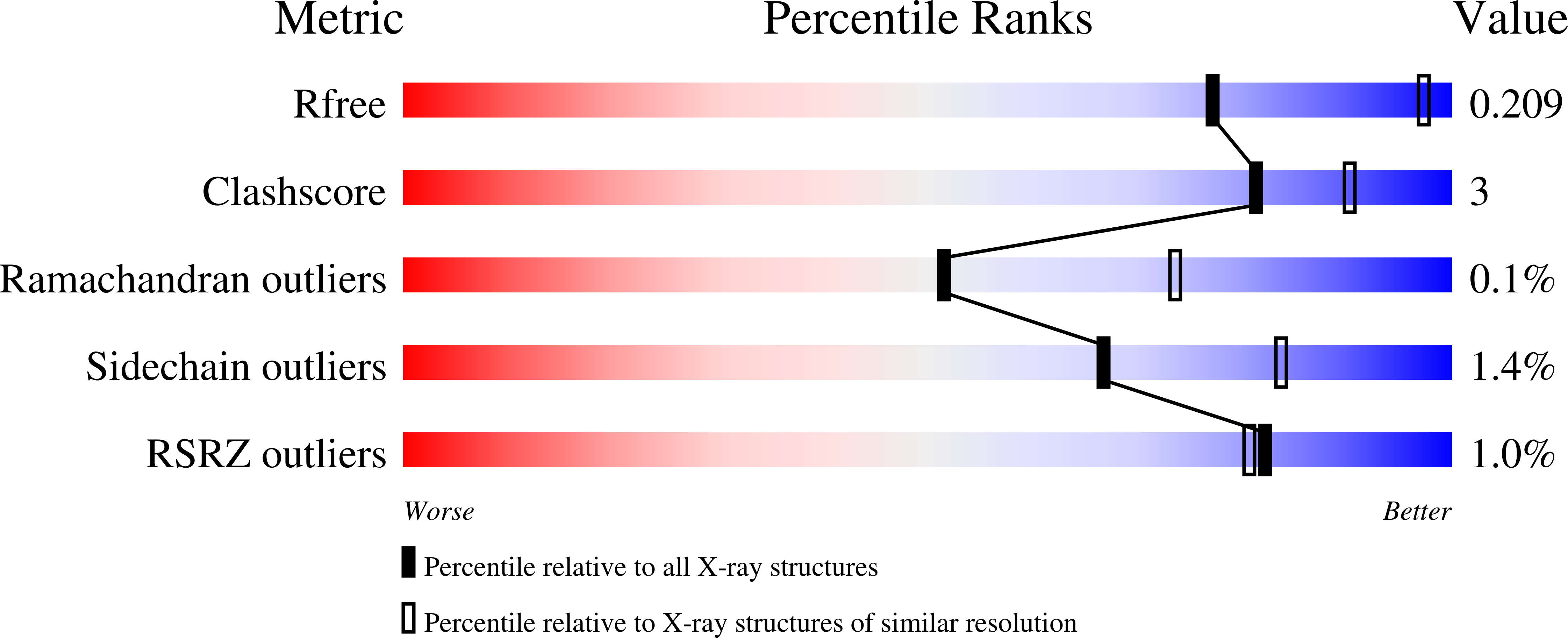
    3DD1, 3DDS

  • PubMed Abstract: 

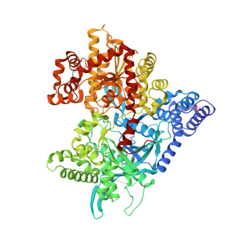
    Key binding interactions of the anthranilimide based glycogen phosphorylase a (GPa) inhibitor 2 from X-ray crystallography studies are described. This series of compounds bind to the AMP site of GP. Using the binding information the core and the phenyl urea moieties were optimized. This work culminated in the identification of compounds with single nanomolar potency as well as in vivo efficacy in a diabetic model.


  • Organizational Affiliation

    Metabolic Center for Excellence in Drug Discovery, GlaxoSmithKline, 5 Moore Drive, PO Box 13398, Research Triangle Park, NC 27705, USA. stephen.a.thomson@gsk.com


Macromolecules
Find similar proteins by:  (by identity cutoff)  |  3D Structure
Entity ID: 1
MoleculeChains Sequence LengthOrganismDetailsImage
Glycogen phosphorylase, liver form
A, B
848Homo sapiensMutation(s): 0 
Gene Names: PYGL
EC: 2.4.1.1
UniProt & NIH Common Fund Data Resources
Find proteins for P06737 (Homo sapiens)
Explore P06737 
Go to UniProtKB:  P06737
PHAROS:  P06737
GTEx:  ENSG00000100504 
Entity Groups  
Sequence Clusters30% Identity50% Identity70% Identity90% Identity95% Identity100% Identity
UniProt GroupP06737
Sequence Annotations
Expand
  • Reference Sequence
Small Molecules
Ligands 6 Unique
IDChains Name / Formula / InChI Key2D Diagram3D Interactions
25D
Query on 25D

Download Ideal Coordinates CCD File 
F [auth A],
O [auth B]
2-cyclohexyl-N-[(3-{[(2,4,6-trimethylphenyl)carbamoyl]amino}naphthalen-2-yl)carbonyl]-D-alanine
C30 H35 N3 O4
ZYHGLTOSKWXGQM-PMERELPUSA-N
PLP
Query on PLP

Download Ideal Coordinates CCD File 
D [auth A],
K [auth B]
PYRIDOXAL-5'-PHOSPHATE
C8 H10 N O6 P
NGVDGCNFYWLIFO-UHFFFAOYSA-N
NBG
Query on NBG

Download Ideal Coordinates CCD File 
C [auth A],
J [auth B]
N-acetyl-beta-D-glucopyranosylamine
C8 H15 N O6
IBONACLSSOLHFU-JAJWTYFOSA-N
MES
Query on MES

Download Ideal Coordinates CCD File 
M [auth B],
N [auth B]
2-(N-MORPHOLINO)-ETHANESULFONIC ACID
C6 H13 N O4 S
SXGZJKUKBWWHRA-UHFFFAOYSA-N
CFF
Query on CFF

Download Ideal Coordinates CCD File 
E [auth A],
L [auth B]
CAFFEINE
C8 H10 N4 O2
RYYVLZVUVIJVGH-UHFFFAOYSA-N
MPD
Query on MPD

Download Ideal Coordinates CCD File 
G [auth A],
H [auth A],
I [auth A],
P [auth B],
Q [auth B]
(4S)-2-METHYL-2,4-PENTANEDIOL
C6 H14 O2
SVTBMSDMJJWYQN-YFKPBYRVSA-N
Modified Residues  1 Unique
IDChains TypeFormula2D DiagramParent
SEP
Query on SEP
A, B
L-PEPTIDE LINKINGC3 H8 N O6 PSER
Binding Affinity Annotations 
IDSourceBinding Affinity
CFF BindingDB:  3DD1 IC50: 6.48e+5 (nM) from 1 assay(s)
25D BindingDB:  3DD1 IC50: min: 10, max: 350 (nM) from 2 assay(s)
Experimental Data & Validation

Experimental Data

  • Method: X-RAY DIFFRACTION
  • Resolution: 2.57 Å
  • R-Value Free: 0.207 
  • R-Value Work: 0.167 
  • R-Value Observed: 0.169 
  • Space Group: P 31
Unit Cell:
Length ( Å )Angle ( ˚ )
a = 124.103α = 90
b = 124.103β = 90
c = 123.222γ = 120
Software Package:
Software NamePurpose
REFMACrefinement
ADSCdata collection
HKL-2000data reduction
HKL-2000data scaling
AMoREphasing

Structure Validation

View Full Validation Report


Ligand Structure Quality Assessment 


Entry History 

Deposition Data

  • Released Date: 2009-04-21 
  • Deposition Author(s): Nolte, R.T.

Revision History  (Full details and data files)

  • Version 1.0: 2009-04-21
    Type: Initial release
  • Version 1.1: 2011-07-13
    Changes: Non-polymer description, Version format compliance
  • Version 1.2: 2020-07-29
    Type: Remediation
    Reason: Carbohydrate remediation
    Changes: Data collection, Database references, Derived calculations, Structure summary
  • Version 1.3: 2023-08-30
    Changes: Data collection, Database references, Refinement description, Structure summary